Loading
PDBj
MenuPDBj@FacebookPDBj@TwitterPDBj@YouTubewwPDB FoundationwwPDB
RCSB PDBPDBeBMRBAdv. SearchSearch help
Search by PDB author
2B7A
DownloadVisualize
BU of 2b7a by Molmil
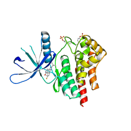
The structural basis of Janus Kinase 2 inhibition by a potent and specific pan-Janus kinase inhibitor
Descriptor: 2-TERT-BUTYL-9-FLUORO-3,6-DIHYDRO-7H-BENZ[H]-IMIDAZ[4,5-F]ISOQUINOLINE-7-ONE, Tyrosine-protein kinase JAK2
Authors:Lucet, I.S, Fantino, E, Styles, M, Bamert, R, Patel, O, Broughton, S.E, Walter, M, Burns, C.J, Treutlein, H, Wilks, A.F, Rossjohn, J.
Deposit date:2005-10-04
Release date:2006-01-10
Last modified:2023-11-15
Method:X-RAY DIFFRACTION (2 Å)
Cite:The structural basis of Janus kinase 2 inhibition by a potent and specific pan-Janus kinase inhibitor.
Blood, 107, 2006
2OGV
DownloadVisualize
BU of 2ogv by Molmil
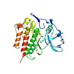
Crystal Structure of the Autoinhibited Human c-Fms Kinase Domain
Descriptor: Macrophage colony-stimulating factor 1 receptor precursor
Authors:Walter, M, Lucet, I.S, Patel, O, Broughton, S.E, Bamert, R, Williams, N.K, Fantino, E, Wilks, A.F, Rossjohn, J.
Deposit date:2007-01-09
Release date:2007-02-06
Last modified:2023-08-30
Method:X-RAY DIFFRACTION (2.7 Å)
Cite:The 2.7 A crystal structure of the autoinhibited human c-Fms kinase domain.
J.Mol.Biol., 367, 2007
5W68
DownloadVisualize
BU of 5w68 by Molmil
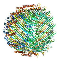
Type II secretin from Enteropathogenic Escherichia coli - GspD
Descriptor: Putative type II secretion protein
Authors:Hay, I.D, Belousoff, M.J, Dunstan, R, Bamert, R, Lithgow, T.
Deposit date:2017-06-16
Release date:2017-11-15
Last modified:2024-03-13
Method:ELECTRON MICROSCOPY (3.3 Å)
Cite:Structure and Membrane Topography of the Vibrio-Type Secretin Complex from the Type 2 Secretion System of Enteropathogenic Escherichia coli.
J. Bacteriol., 200, 2018
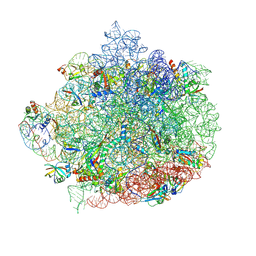
6WQQ
DownloadVisualize
BU of 6wqq by Molmil
Structure of the 50S subunit of the ribosome from Methicillin Resistant Staphylococcus aureus in complex with the antibiotic, radezolid
Descriptor: 23S rRNA, 50S ribosomal protein L13, 50S ribosomal protein L14, ...
Authors:Belousoff, M.J.
Deposit date:2020-04-29
Release date:2020-06-03
Last modified:2020-12-16
Method:ELECTRON MICROSCOPY (3.1 Å)
Cite:Characterization of the Core Ribosomal Binding Region for the Oxazolidone Family of Antibiotics Using Cryo-EM.
Acs Pharmacol Transl Sci, 3, 2020
6WRU
DownloadVisualize
BU of 6wru by Molmil
Structure of the 50S subunit of the ribosome from Methicillin Resistant Staphylococcus aureus in complex with an isomer of the tedizolid
Descriptor: 23S rRNA, 50S ribosomal protein L13, 50S ribosomal protein L14, ...
Authors:Belousoff, M.J.
Deposit date:2020-04-30
Release date:2020-06-03
Last modified:2020-12-16
Method:ELECTRON MICROSCOPY (3.1 Å)
Cite:Characterization of the Core Ribosomal Binding Region for the Oxazolidone Family of Antibiotics Using Cryo-EM.
Acs Pharmacol Transl Sci, 3, 2020
6WRS
DownloadVisualize
BU of 6wrs by Molmil
Structure of the 50S subunit of the ribosome from Methicillin Resistant Staphylococcus aureus in complex with the antibiotic, tedizolid
Descriptor: 23S rRNA, 50S ribosomal protein L13, 50S ribosomal protein L14, ...
Authors:Belousoff, M.J.
Deposit date:2020-04-30
Release date:2020-06-03
Last modified:2020-12-16
Method:ELECTRON MICROSCOPY (3.2 Å)
Cite:Characterization of the Core Ribosomal Binding Region for the Oxazolidone Family of Antibiotics Using Cryo-EM.
Acs Pharmacol Transl Sci, 3, 2020
6WQN
DownloadVisualize
BU of 6wqn by Molmil
Structure of the 50S subunit of the ribosome from Methicillin Resistant Staphylococcus aureus in complex with the antibiotic, contezolid
Descriptor: 23S rRNA, 50S ribosomal protein L13, 50S ribosomal protein L14, ...
Authors:Belousoff, M.J.
Deposit date:2020-04-29
Release date:2020-06-03
Last modified:2020-12-16
Method:ELECTRON MICROSCOPY (2.9 Å)
Cite:Characterization of the Core Ribosomal Binding Region for the Oxazolidone Family of Antibiotics Using Cryo-EM.
Acs Pharmacol Transl Sci, 3, 2020

218853

PDB entries from 2024-04-24

PDB statisticsPDBj update infoContact PDBjnumon