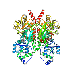
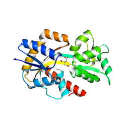
3MPK
 
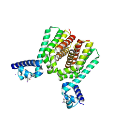
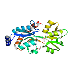 | | Crystal Structure of Bordetella pertussis BvgS periplasmic VFT2 domain | | Descriptor: | ACETATE ION, GLYCEROL, Virulence sensor protein bvgS | | Authors: | Herrou, J, Bompard, C, Wintjens, R, Dupre, E, Willery, E, Villeret, V, Locht, C, Antoine, R, Jacob-Dubuisson, F. | | Deposit date: | 2010-04-27 | | Release date: | 2010-10-06 | | Last modified: | 2024-02-21 | | Method: | X-RAY DIFFRACTION (2.04 Å) | | Cite: | Periplasmic domain of the sensor-kinase BvgS reveals a new paradigm for the Venus flytrap mechanism.
Proc.Natl.Acad.Sci.USA, 107, 2010
|
|
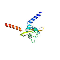
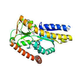
2DVZ
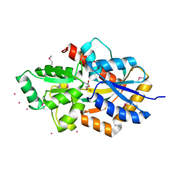 | | Structure of a periplasmic transporter | | Descriptor: | CADMIUM ION, GLUTAMIC ACID, Putative exported protein | | Authors: | Huvent, I, Belrhali, H, Antoine, R, Bompard, C, Locht, C, Jacob-Dubuisson, F, Villeret, V. | | Deposit date: | 2006-08-01 | | Release date: | 2006-11-07 | | Last modified: | 2011-07-13 | | Method: | X-RAY DIFFRACTION (2.3 Å) | | Cite: | Structural analysis of Bordetella pertussis BugE solute receptor in a bound conformation
ACTA CRYSTALLOGR.,SECT.D, 62, 2006
|
|
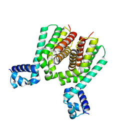
3MPL
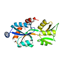 | | Crystal Structure of Bordetella pertussis BvgS VFT2 domain (Double Mutant F375E/Q461E) | | Descriptor: | 1,2-ETHANEDIOL, Virulence sensor protein bvgS | | Authors: | Herrou, J, Bompard, C, Wintjens, R, Dupre, E, Willery, E, Villeret, V, Locht, C, Antoine, R, Jacob-Dubuisson, F. | | Deposit date: | 2010-04-27 | | Release date: | 2010-10-06 | | Last modified: | 2023-09-06 | | Method: | X-RAY DIFFRACTION (2.1 Å) | | Cite: | Periplasmic domain of the sensor-kinase BvgS reveals a new paradigm for the Venus flytrap mechanism.
Proc.Natl.Acad.Sci.USA, 107, 2010
|
|
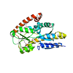
2F5X
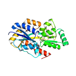 | | Structure of periplasmic binding protein BugD | | Descriptor: | ASPARTIC ACID, BugD | | Authors: | Huvent, I, Belrhali, H, Antoine, R, Bompard, C, Jacob-Dubuisson, F, Villeret, V. | | Deposit date: | 2005-11-28 | | Release date: | 2006-01-24 | | Last modified: | 2011-07-13 | | Method: | X-RAY DIFFRACTION (1.72 Å) | | Cite: | Crystal Structure of Bordetella pertussis BugD Solute Receptor Unveils the Basis of Ligand Binding in a New Family of Periplasmic Binding Proteins
J.Mol.Biol., 356, 2006
|
|
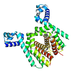
4Q0C
 | | 3.1 A resolution crystal structure of the B. pertussis BvgS periplasmic domain | | Descriptor: | Virulence sensor protein BvgS | | Authors: | Dupre, E, Herrou, J, Lensink, M.F, Wintjens, R, Lebedev, A, Crosson, S, Villeret, V, Locht, C, Antoine, R, Jacob-Dubuisson, F. | | Deposit date: | 2014-04-01 | | Release date: | 2015-02-11 | | Last modified: | 2023-09-20 | | Method: | X-RAY DIFFRACTION (3.1 Å) | | Cite: | Virulence Regulation with Venus Flytrap Domains: Structure and Function of the Periplasmic Moiety of the Sensor-Kinase BvgS.
Plos Pathog., 11, 2015
|
|
6ZJ8
 | |
5ICJ
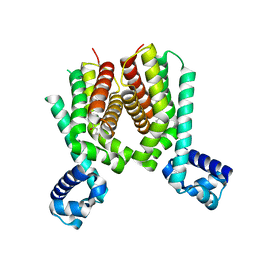 | | Crystal structure of the Mycobacterium tuberculosis transcriptional repressor EthR2 in complex with BDM41420 | | Descriptor: | 4,4,4-trifluoro-1-(3-phenyl-1-oxa-2,8-diazaspiro[4.5]dec-2-en-8-yl)butan-1-one, Probable transcriptional regulatory protein | | Authors: | Wohlkonig, A, Remaut, H, Tanina, A, Meyer, F, Willand, N, Baulard, A.R, Wintjens, R. | | Deposit date: | 2016-02-23 | | Release date: | 2017-04-26 | | Last modified: | 2024-01-10 | | Method: | X-RAY DIFFRACTION (2.4 Å) | | Cite: | Reversion of antibiotic resistance in Mycobacterium tuberculosis by spiroisoxazoline SMARt-420.
Science, 355, 2017
|
|
5N1I
 | |
5N1C
 | |
5N7O
 | | EthR2 in complex with SMARt-420 compound | | Descriptor: | 4,4,4-trifluoro-1-(3-phenyl-1-oxa-2,8-diazaspiro[4.5]dec-2-en-8-yl)butan-1-one, Probable transcriptional regulatory protein | | Authors: | Wohlkonig, A, Wintjens, R. | | Deposit date: | 2017-02-21 | | Release date: | 2017-04-26 | | Last modified: | 2024-01-17 | | Method: | X-RAY DIFFRACTION (2.3 Å) | | Cite: | Structural analysis of the interaction between spiroisoxazoline SMARt-420 and the Mycobacterium tuberculosis repressor EthR2.
Biochem. Biophys. Res. Commun., 487, 2017
|
|
2PFZ
 | |
2PFY
 | |
2QPQ
 | | Structure of Bug27 from Bordetella pertussis | | Descriptor: | CITRIC ACID, protein Bug27 | | Authors: | Herrou, J, Bompard, C. | | Deposit date: | 2007-07-25 | | Release date: | 2007-08-21 | | Last modified: | 2011-07-13 | | Method: | X-RAY DIFFRACTION (1.92 Å) | | Cite: | Structure-based mechanism of ligand binding for periplasmic solute-binding protein of the Bug family.
J.Mol.Biol., 373, 2007
|
|


