3F81
 
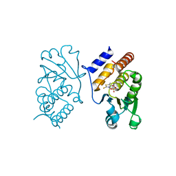 | | Interaction of VHR with SA3 | | Descriptor: | 2-[(5~{E})-5-[[3-[4-(2-fluoranylphenoxy)phenyl]-1-phenyl-pyrazol-4-yl]methylidene]-4-oxidanylidene-2-sulfanylidene-1,3-thiazolidin-3-yl]ethanesulfonic acid, Dual specificity protein phosphatase 3 | | Authors: | Wu, S, Mutelin, T, Tautz, L. | | Deposit date: | 2008-11-11 | | Release date: | 2009-11-10 | | Last modified: | 2023-09-06 | | Method: | X-RAY DIFFRACTION (1.9 Å) | | Cite: | Multidentate small-molecule inhibitors of vaccinia H1-related (VHR) phosphatase decrease proliferation of cervix cancer cells.
J.Med.Chem., 52, 2009
|
|
2F46
 | |
1ZC0
 | |
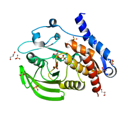
6YID
 | | Crystal structure of ULK2 in complex with SBI-0206965 | | Descriptor: | 2-({5-bromo-2-[(3,4,5-trimethoxyphenyl)amino]pyrimidin-4-yl}oxy)-N-methylbenzene-1-carboximidic acid, Serine/threonine-protein kinase ULK2 | | Authors: | Chaikuad, A, Ren, H, Bakas, N.A, Lambert, L.J, Cosford, N.D.P, Knapp, S, Structural Genomics Consortium (SGC) | | Deposit date: | 2020-04-01 | | Release date: | 2020-06-17 | | Last modified: | 2024-01-24 | | Method: | X-RAY DIFFRACTION (2.7 Å) | | Cite: | Design, Synthesis, and Characterization of an Orally Active Dual-Specific ULK1/2 Autophagy Inhibitor that Synergizes with the PARP Inhibitor Olaparib for the Treatment of Triple-Negative Breast Cancer.
J.Med.Chem., 63, 2020
|
|
3O4S
 | |
3O4T
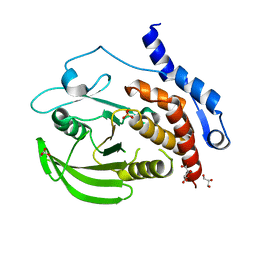 | |
3O4U
 | | Crystal Structure of HePTP with an Atypically Open WPD Loop | | Descriptor: | GLYCEROL, L(+)-TARTARIC ACID, S,R MESO-TARTARIC ACID, ... | | Authors: | Critton, D.A, Page, R. | | Deposit date: | 2010-07-27 | | Release date: | 2010-11-24 | | Last modified: | 2023-09-06 | | Method: | X-RAY DIFFRACTION (2.25 Å) | | Cite: | Visualizing active-site dynamics in single crystals of HePTP: opening of the WPD loop involves coordinated movement of the E loop.
J.Mol.Biol., 405, 2011
|
|


