Loading
PDBj
MenuPDBj@FacebookPDBj@TwitterPDBj@YouTubewwPDB FoundationwwPDB
RCSB PDBPDBeBMRBAdv. SearchSearch help
Search by PDB author
4Z37
DownloadVisualize
BU of 4z37 by Molmil
Structure of the ketosynthase of module 2 of C0ZGQ5 (trans-AT PKS) from Brevibacillus brevis
Descriptor: Putative mixed polyketide synthase/non-ribosomal peptide synthetase
Authors:Martin, S.F, Jakob, R.P, Herbst, D.A, Maier, T.
Deposit date:2015-03-31
Release date:2016-04-13
Last modified:2024-01-10
Method:X-RAY DIFFRACTION (1.998 Å)
Cite:The structural organization of trans-AT polyketide synthases: ketoacyl synthase and trans-acting enoyl reductase
To Be Published
6WM1
DownloadVisualize
BU of 6wm1 by Molmil
Crystal structure of the Grb2 SH2 domain in complex with a tripeptide: Ac-pY-Ac6c-N-phenylpropyl
Descriptor: ACE-PTR-02K-ASN-PRA, CALCIUM ION, CHLORIDE ION, ...
Authors:Martin, S.F, Clements, J.H.
Deposit date:2020-04-20
Release date:2020-09-02
Last modified:2023-11-15
Method:X-RAY DIFFRACTION (1.8 Å)
Cite:Some thermodynamic effects of varying nonpolar surfaces in protein-ligand interactions.
Eur.J.Med.Chem., 208, 2020
6WO2
DownloadVisualize
BU of 6wo2 by Molmil
Crystal Structure of the Grb2 SH2 Domain in Complex with a Tripeptide: Ac-pY-Ac6c-N-isohexyl
Descriptor: ACE-PTR-02K-ASN-U67, CALCIUM ION, Growth factor receptor-bound protein 2, ...
Authors:Martin, S.F, Clements, J.H.
Deposit date:2020-04-24
Release date:2020-09-02
Last modified:2023-11-15
Method:X-RAY DIFFRACTION (2 Å)
Cite:Some thermodynamic effects of varying nonpolar surfaces in protein-ligand interactions.
Eur.J.Med.Chem., 208, 2020
4Z38
DownloadVisualize
BU of 4z38 by Molmil
Crystal structure of enoyl reductase domain of MlnA from the macrolactin biosynthesis cluster from Bacillus amyloliquefaciens
Descriptor: FLAVIN MONONUCLEOTIDE, MlnA
Authors:Jakob, R.P, Martin, S.F, Herbst, D.A, Maier, T.
Deposit date:2015-03-31
Release date:2016-04-13
Last modified:2024-01-10
Method:X-RAY DIFFRACTION (2.8 Å)
Cite:The structural organization of trans-AT polyketide synthases: ketoacyl synthase and trans-acting enoyl reductase
To Be Published
2FFZ
DownloadVisualize
BU of 2ffz by Molmil
Structural Studies Examining the Substrate Specificity Profiles of PC-PLCBc Protein Variants
Descriptor: Phospholipase C, ZINC ION
Authors:Benfield, A.B, Antikainen, N.M, Martin, S.F.
Deposit date:2005-12-20
Release date:2006-03-28
Last modified:2024-02-14
Method:X-RAY DIFFRACTION (2.05 Å)
Cite:Structural studies examining the substrate specificity profiles of PC-PLC(Bc) protein variants.
Arch.Biochem.Biophys., 460, 2007
2FGN
DownloadVisualize
BU of 2fgn by Molmil
Structural Studies Examining the Substrate Specificity Profiles of PC-PLCBc Protein Variants
Descriptor: Phospholipase C, ZINC ION
Authors:Benfield, A.P, Martin, S.F.
Deposit date:2005-12-22
Release date:2006-03-28
Last modified:2024-02-14
Method:X-RAY DIFFRACTION (2.04 Å)
Cite:Structural studies examining the substrate specificity profiles of PC-PLC(Bc) protein variants.
Arch.Biochem.Biophys., 460, 2007
1IS0
DownloadVisualize
BU of 1is0 by Molmil
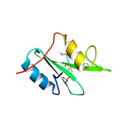
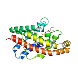
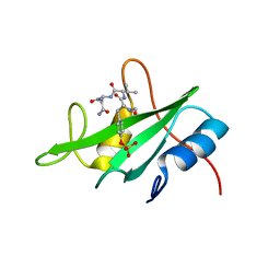
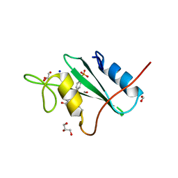
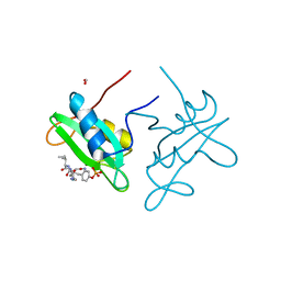
Crystal Structure of a Complex of the Src SH2 Domain with Conformationally Constrained Peptide Inhibitor
Descriptor: AY0 GLU GLU ILE peptide, Tyrosine-protein kinase transforming protein SRC
Authors:Davidson, J.P, Lubman, O, Rose, T, Waksman, G, Martin, S.F.
Deposit date:2001-11-02
Release date:2002-02-06
Last modified:2023-12-27
Method:X-RAY DIFFRACTION (1.9 Å)
Cite:Calorimetric and structural studies of 1,2,3-trisubstituted cyclopropanes as conformationally constrained peptide inhibitors of Src SH2 domain binding.
J.Am.Chem.Soc., 124, 2002
1P6D
DownloadVisualize
BU of 1p6d by Molmil
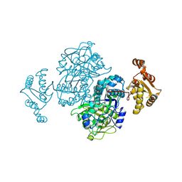
STRUCTURE OF THE D55N MUTANT OF PHOSPHOLIPASE C FROM BACILLUS CEREUS IN COMPLEX WITH (3S)-3,4,DI-N-HEXANOYLOXYBUTYL-1-PHOSPHOCHOLINE
Descriptor: (3S)-3,4-DI-N-HEXANOYLOXYBUTYL-1-PHOSPHOCHOLINE, PHOSPHOLIPASE C, ZINC ION
Authors:Antikainen, N.M, Monzingo, A.F, Franklin, C.L, Robertus, J.D, Martin, S.F.
Deposit date:2003-04-29
Release date:2003-09-30
Last modified:2023-08-16
Method:X-RAY DIFFRACTION (2 Å)
Cite:Using X-ray crystallography of the Asp55Asn mutant of the phosphatidylcholine-preferring phospholipase C from Bacillus cereus to support the mechanistic role of Asp55 as the general base.
Arch.Biochem.Biophys., 417, 2003
1P5X
DownloadVisualize
BU of 1p5x by Molmil
STRUCTURE OF THE D55N MUTANT OF PHOSPHOLIPASE C FROM BACILLUS CEREUS
Descriptor: Phospholipase C, ZINC ION
Authors:Antikainen, N.M, Monzingo, A.F, Franklin, C.L, Robertus, J.D, Martin, S.F.
Deposit date:2003-04-28
Release date:2003-09-30
Last modified:2023-08-16
Method:X-RAY DIFFRACTION (2 Å)
Cite:Using X-ray crystallography of the Asp55Asn mutant of the phosphatidylcholine-preferring phospholipase C from Bacillus cereus to support the mechanistic role of Asp55 as the general base.
Arch.Biochem.Biophys., 417, 2003
1P6E
DownloadVisualize
BU of 1p6e by Molmil
STRUCTURE OF THE D55N MUTANT OF PHOSPHOLIPASE C FROM BACILLUS CEREUS IN COMPLEX WITH 1,2-DI-N-PENTANOYL-SN-GLYCERO-3-DITHIOPHOSPHOCHOLINE
Descriptor: 1,2-DI-N-PENTANOYL-SN-GLYCERO-3-DITHIOPHOSPHOCHOLINE, Phospholipase C, ZINC ION
Authors:Antikainen, N.M, Monzingo, A.F, Franklin, C.L, Robertus, J.D, Martin, S.F.
Deposit date:2003-04-29
Release date:2003-09-30
Last modified:2023-08-16
Method:X-RAY DIFFRACTION (2.3 Å)
Cite:Using X-ray crystallography of the Asp55Asn mutant of the phosphatidylcholine-preferring phospholipase C from Bacillus cereus to support the mechanistic role of Asp55 as the general base.
Arch.Biochem.Biophys., 417, 2003
3C7I
DownloadVisualize
BU of 3c7i by Molmil
X-RAY crystal structure of the complex between the grb2-sh2 domain and a flexible ligand, FPTVN.
Descriptor: FORMIC ACID, Growth factor receptor-bound protein 2, N-{(2R)-4-(methylamino)-4-oxo-2-[4-(phosphonooxy)benzyl]butanoyl}-L-valyl-L-aspartamide
Authors:Benfield, A.P, Martin, S.F.
Deposit date:2008-02-07
Release date:2008-08-12
Last modified:2024-02-21
Method:X-RAY DIFFRACTION (1.7 Å)
Cite:Ligand Preorganization May be Accompanied by Entropic Penalties in Protein-Ligand Interactions.
Angew.Chem.Int.Ed.Engl., 118, 2006
2H5K
DownloadVisualize
BU of 2h5k by Molmil
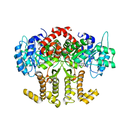
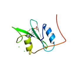
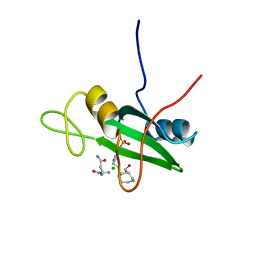
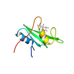
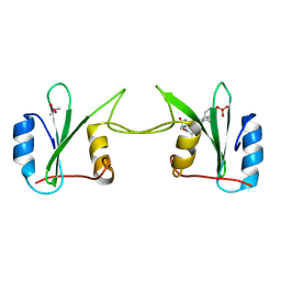
Crystal Structure of Complex Between the Domain-Swapped Dimeric Grb2 SH2 Domain and Shc-Derived Ligand, Ac-NH-pTyr-Val-Asn-NH2
Descriptor: CACODYLATE ION, Growth factor receptor-bound protein 2, Shc-Derived Ligand
Authors:Benfield, A.P, Whiddon, B.B, Martin, S.F.
Deposit date:2006-05-26
Release date:2006-08-15
Last modified:2017-10-18
Method:X-RAY DIFFRACTION (3.25 Å)
Cite:Structural and energetic aspects of Grb2-SH2 domain-swapping.
Arch.Biochem.Biophys., 462, 2007
2H46
DownloadVisualize
BU of 2h46 by Molmil
Native domain-swapped dimer crystal structure of the Grb2 SH2 domain
Descriptor: GLYCEROL, Growth Receptor Binding Protein 2
Authors:Benfield, A.P, Martin, S.F, Whiddon, B.B.
Deposit date:2006-05-23
Release date:2006-06-06
Last modified:2024-02-14
Method:X-RAY DIFFRACTION (1.9 Å)
Cite:Structural and energetic aspects of Grb2-SH2 domain-swapping.
Arch.Biochem.Biophys., 462, 2007
2HUC
DownloadVisualize
BU of 2huc by Molmil
Structural Studies Examining the Substrate Specificity Profiles of PC-PLCBc Protein Variants
Descriptor: Phospholipase C, ZINC ION
Authors:Benfield, A.P, Martin, S.F, Antikainen, N.M.
Deposit date:2006-07-26
Release date:2006-08-08
Last modified:2024-02-14
Method:X-RAY DIFFRACTION (1.9 Å)
Cite:Structural studies examining the substrate specificity profiles of PC-PLC(Bc) protein variants.
Arch.Biochem.Biophys., 460, 2007
2HUW
DownloadVisualize
BU of 2huw by Molmil
X-ray crystal structure of the Grb2 SH2 domain complexed to a constrained and cyclopropane-derived ligand
Descriptor: Growth factor receptor-bound protein 2, N-({(1R,2R,3S)-2-(methylcarbamoyl)-3-[4-(phosphonooxy)phenyl]cyclopropyl}carbonyl)-L-valyl-L-aspartamide, PHOSPHATE ION
Authors:Benfield, A.P, Martin, S.F.
Deposit date:2006-07-27
Release date:2006-08-15
Last modified:2024-02-14
Method:X-RAY DIFFRACTION (1.9 Å)
Cite:Ligand Preorganization May Be Accompanied by Entropic Penalties in Protein-Ligand Interactions.
Angew.Chem.Int.Ed.Engl., 45, 2006
3N84
DownloadVisualize
BU of 3n84 by Molmil
Crystal Structure of the Grb2 SH2 Domain in Complex with a 23-Membered Macrocyclic Ligand Having the Sequence pYVNVP
Descriptor: 23-membered peptide-like macrocyclic ligand, CHLORIDE ION, GLYCEROL, ...
Authors:Clements, J.H, Martin, S.F.
Deposit date:2010-05-27
Release date:2011-01-12
Last modified:2023-11-15
Method:X-RAY DIFFRACTION (2 Å)
Cite:Thermodynamic and Structural Effects of Macrocyclization as a Constraining Method in Protein-Ligand Interactions.
ACS MED.CHEM.LETT., 1, 2010
3N8M
DownloadVisualize
BU of 3n8m by Molmil
Crystal Structure of the Grb2 SH2 Domain in Complex with An Acyclic Ligand Having the Sequence pYVNVP
Descriptor: GLYCEROL, Growth factor receptor-bound protein 2, PEPTIDE
Authors:Whiddon, B.B, Clements, J.H, Martin, S.F.
Deposit date:2010-05-28
Release date:2010-12-15
Last modified:2023-11-22
Method:X-RAY DIFFRACTION (2 Å)
Cite:Thermodynamic and Structural Effects of Macrocyclization as a Constraining Method in Protein-Ligand Interactions.
ACS MED.CHEM.LETT., 1, 2010
3N7Y
DownloadVisualize
BU of 3n7y by Molmil
Crystal Structure of the Grb2 SH2 Domain in Complex with a 20-Membered Macrocyclic Ligand Having the Sequence pYVNV
Descriptor: 20-membered peptide-like macrocyclic ligand, Growth factor receptor-bound protein 2
Authors:Whiddon, B.B, Clements, J.H, Martin, S.F.
Deposit date:2010-05-27
Release date:2011-01-12
Last modified:2023-11-15
Method:X-RAY DIFFRACTION (2.02 Å)
Cite:Thermodynamic and Structural Effects of Macrocyclization as a Constraining Method in Protein-Ligand Interactions.
ACS MED.CHEM.LETT., 1, 2010
3OV1
DownloadVisualize
BU of 3ov1 by Molmil
Crystal Structure of the Grb2 SH2 Domain in Complex with a pYXN-Derived Tripeptide
Descriptor: FORMIC ACID, GLYCEROL, Growth factor receptor-bound protein 2, ...
Authors:Clements, J.H, Martin, S.F.
Deposit date:2010-09-15
Release date:2011-11-02
Last modified:2023-12-06
Method:X-RAY DIFFRACTION (1.6 Å)
Cite:Protein-ligand interactions: thermodynamic effects associated with increasing nonpolar surface area.
J.Am.Chem.Soc., 133, 2011
3OVE
DownloadVisualize
BU of 3ove by Molmil
Crystal Structure of the Grb2 SH2 Domain in Complex with a pYXN-Derived Tripeptide
Descriptor: CHLORIDE ION, Growth factor receptor-bound protein 2, PYAC7CN
Authors:Clements, J.H, Martin, S.F.
Deposit date:2010-09-16
Release date:2011-11-02
Last modified:2023-12-06
Method:X-RAY DIFFRACTION (1.82 Å)
Cite:Protein-ligand interactions: thermodynamic effects associated with increasing nonpolar surface area.
J.Am.Chem.Soc., 133, 2011
4P9V
DownloadVisualize
BU of 4p9v by Molmil
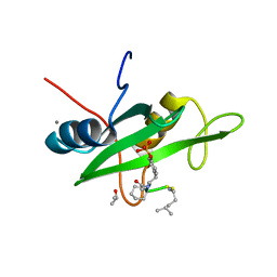
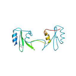
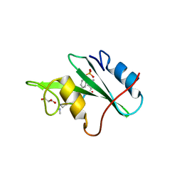
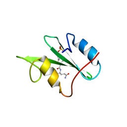
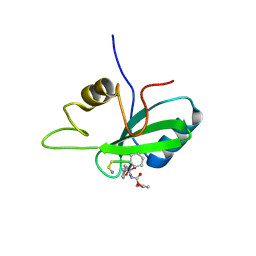
Grb2 SH2 complexed with a pTyr-Ac6cN-Asn tripeptide
Descriptor: CHLORIDE ION, Growth factor receptor-bound protein 2, PHQ-PTR-02K-ASN-NH2
Authors:Clements, J.H, Martin, S.F.
Deposit date:2014-04-06
Release date:2014-06-18
Last modified:2023-11-15
Method:X-RAY DIFFRACTION (1.64 Å)
Cite:Protein-ligand interactions: Probing the energetics of a putative cation-pi interaction.
Bioorg.Med.Chem.Lett., 24, 2014
4P9Z
DownloadVisualize
BU of 4p9z by Molmil
Grb2 SH2 complexed with a pTyr-Ac6c-Asn tripeptide
Descriptor: ACETATE ION, CHLORIDE ION, GLYCEROL, ...
Authors:Clements, J.H, Martin, S.F.
Deposit date:2014-04-06
Release date:2014-06-18
Last modified:2023-12-27
Method:X-RAY DIFFRACTION (1.8 Å)
Cite:Protein-ligand interactions: Probing the energetics of a putative cation-pi interaction.
Bioorg.Med.Chem.Lett., 24, 2014
3IN8
DownloadVisualize
BU of 3in8 by Molmil
Crystal Structure of the Grb2 SH2 Domain in Complex with a Flexible Ac-pTyr-Ile-Asn-NH2 Tripeptide Mimic
Descriptor: FORMIC ACID, Growth factor receptor-bound protein 2, N-{(2S)-4-(methylamino)-4-oxo-2-[4-(phosphonooxy)benzyl]butanoyl}-L-isoleucyl-L-aspartamide
Authors:Benfield, A.P, Clements, J.H.
Deposit date:2009-08-11
Release date:2009-11-17
Last modified:2023-09-06
Method:X-RAY DIFFRACTION (1.7 Å)
Cite:Thermodynamic and Structural Effects of Conformational Constraints in Protein-Ligand Interactions. Entropic Paradoxy Associated with Ligand Preorganization.
J.Am.Chem.Soc., 131, 2009
3IMJ
DownloadVisualize
BU of 3imj by Molmil
Crystal Structure of the Grb2 SH2 Domain in Complex with a Cyclopropyl-constrained Ac-pTyr-Ile-Asn-NH2 Tripeptide Mimic
Descriptor: (1R,2S,3R,1S',1S")Phosphoric acid mono(4-{2-[1-(1,2-dicarbamoylethylcarbamoyl)-3-carbamoylpropylcarbamoyl]-3-methylcarbamoylcyclopropyl}phenyl) ester, Growth factor receptor-bound protein 2
Authors:Clements, J.H.
Deposit date:2009-08-10
Release date:2009-11-17
Last modified:2023-09-06
Method:X-RAY DIFFRACTION (2.02 Å)
Cite:Thermodynamic and Structural Effects of Conformational Constraints in Protein-Ligand Interactions. Entropic Paradoxy Associated with Ligand Preorganization.
J.Am.Chem.Soc., 131, 2009
3IN7
DownloadVisualize
BU of 3in7 by Molmil
Crystal Structure of the Grb2 SH2 Domain in Complex with a Cyclopropyl-constrained Ac-pY-Q-N-NH2 Tripeptide Mimic
Descriptor: Growth factor receptor-bound protein 2, N~2~-({(1R,2R,3S)-2-(methylcarbamoyl)-3-[4-(phosphonooxy)phenyl]cyclopropyl}carbonyl)-D-glutaminyl-D-aspartamide
Authors:Clements, J.H.
Deposit date:2009-08-11
Release date:2009-11-17
Last modified:2023-09-06
Method:X-RAY DIFFRACTION (2 Å)
Cite:Thermodynamic and Structural Effects of Conformational Constraints in Protein-Ligand Interactions. Entropic Paradoxy Associated with Ligand Preorganization.
J.Am.Chem.Soc., 131, 2009


12>

218500

PDB entries from 2024-04-17

PDB statisticsPDBj update infoContact PDBjnumon