6MWS
 
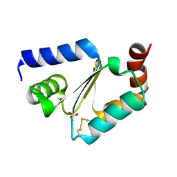 | |
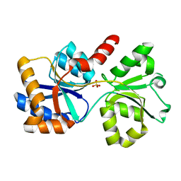
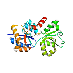
7MQZ
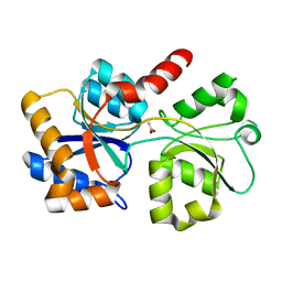
 | | Cytochrome c oxidase assembly factor 7 | | Descriptor: | Cytochrome c oxidase assembly factor 7 | | Authors: | Maghool, S, Maher, M.J. | | Deposit date: | 2021-05-07 | | Release date: | 2022-03-16 | | Last modified: | 2023-10-18 | | Method: | X-RAY DIFFRACTION (2.39 Å) | | Cite: | Mitochondrial COA7 is a heme-binding protein with disulfide reductase activity, which acts in the early stages of complex IV assembly.
Proc.Natl.Acad.Sci.USA, 119, 2022
|
|
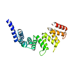
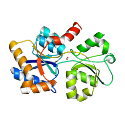
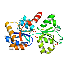
6PCE
 | | Human Coa6 | | Descriptor: | Cytochrome c oxidase assembly factor 6 homolog, SULFATE ION | | Authors: | Maher, M.J, Maghool, S. | | Deposit date: | 2019-06-17 | | Release date: | 2019-10-02 | | Last modified: | 2020-01-01 | | Method: | X-RAY DIFFRACTION (1.65 Å) | | Cite: | Structural and functional characterization of the mitochondrial complex IV assembly factor Coa6.
Life Sci Alliance, 2, 2019
|
|
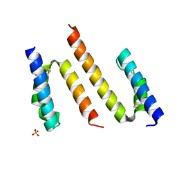
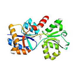
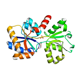
6PCF
 | | Human Coa6: W59C mutant protein | | Descriptor: | Cytochrome c oxidase assembly factor 6 homolog | | Authors: | Maher, M.J, Maghool, S. | | Deposit date: | 2019-06-17 | | Release date: | 2019-10-02 | | Last modified: | 2023-10-11 | | Method: | X-RAY DIFFRACTION (2.2 Å) | | Cite: | Structural and functional characterization of the mitochondrial complex IV assembly factor Coa6.
Life Sci Alliance, 2, 2019
|
|
7L22
 | |
6XAB
 | |
6XAD
 | |
6X9G
 | |
6XL2
 | |
6X8W
 | |
6X6B
 | |


