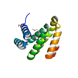
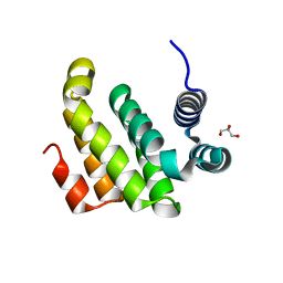
Loading
PDBj
MenuPDBj@FacebookPDBj@TwitterPDBj@YouTubewwPDB FoundationwwPDB
RCSB PDBPDBeBMRBAdv. SearchSearch help
Search by PDB author
1PUF
DownloadVisualize
BU of 1puf by Molmil
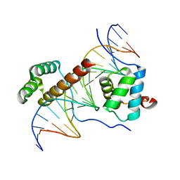
Crystal Structure of HoxA9 and Pbx1 homeodomains bound to DNA
Descriptor: 5'-D(*AP*CP*TP*CP*TP*AP*TP*GP*AP*TP*TP*TP*AP*CP*GP*AP*CP*GP*CP*T)-3', 5'-D(*TP*AP*GP*CP*GP*TP*CP*GP*TP*AP*AP*AP*TP*CP*AP*TP*AP*GP*AP*G)-3', Homeobox protein Hox-A9, ...
Authors:Laronde-Leblanc, N.A, Wolberger, C.
Deposit date:2003-06-24
Release date:2003-09-02
Last modified:2023-08-16
Method:X-RAY DIFFRACTION (1.9 Å)
Cite:STRUCTURE OF HOXA9 AND PBX1 BOUND TO DNA: HOX HEXAPEPTIDE AND DNA RECOGNITION ANTERIOR TO POSTERIOR
Genes Dev., 17, 2003
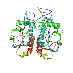
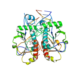
3DLA
DownloadVisualize
BU of 3dla by Molmil
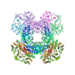
X-ray crystal structure of glutamine-dependent NAD+ synthetase from Mycobacterium tuberculosis bound to NaAD+ and DON
Descriptor: 5-OXO-L-NORLEUCINE, GLYCEROL, Glutamine-dependent NAD(+) synthetase, ...
Authors:LaRonde-LeBlanc, N.A, Resto, M, Gerratana, B.
Deposit date:2008-06-26
Release date:2009-03-10
Last modified:2019-10-23
Method:X-RAY DIFFRACTION (2.35 Å)
Cite:Regulation of active site coupling in glutamine-dependent NAD(+) synthetase.
Nat.Struct.Mol.Biol., 16, 2009
3O7B
DownloadVisualize
BU of 3o7b by Molmil
Crystal structure of Archaeoglobus Fulgidus Nep1 bound to S-adenosylhomocysteine
Descriptor: Ribosome biogenesis Nep1 RNA methyltransferase, S-ADENOSYL-L-HOMOCYSTEINE, TYROSINE
Authors:Thomas, S.R, LaRonde-LeBlanc, N.
Deposit date:2010-07-30
Release date:2011-01-19
Last modified:2017-11-08
Method:X-RAY DIFFRACTION (1.45 Å)
Cite:Structural insight into the functional mechanism of Nep1/Emg1 N1-specific pseudouridine methyltransferase in ribosome biogenesis.
Nucleic Acids Res., 39, 2011
3OII
DownloadVisualize
BU of 3oii by Molmil
Crystal structure of Saccharomyces Cerevisiae Nep1/Emg1 bound to S-adenosylhomocysteine
Descriptor: Essential for mitotic growth 1, GLYCEROL, S-ADENOSYL-L-HOMOCYSTEINE
Authors:Thomas, S.R, Szyk, A, LaRonde-LeBlanc, N.
Deposit date:2010-08-19
Release date:2010-12-01
Last modified:2023-09-06
Method:X-RAY DIFFRACTION (1.85 Å)
Cite:Structural insight into the functional mechanism of Nep1/Emg1 N1-specific pseudouridine methyltransferase in ribosome biogenesis.
Nucleic Acids Res., 39, 2011
3OIN
DownloadVisualize
BU of 3oin by Molmil
Crystal structure of Saccharomyces cerevisiae Nep1/Emg1 bound to S-adenosylhomocysteine and 1 molecule of cognate RNA
Descriptor: 5'-R(*GP*GP*GP*CP*UP*UP*CP*AP*AP*CP*GP*CP*CP*C)-3', CHLORIDE ION, GLYCEROL, ...
Authors:Thomas, S.R, LaRonde-LeBlanc, N.
Deposit date:2010-08-19
Release date:2010-12-01
Last modified:2023-09-06
Method:X-RAY DIFFRACTION (1.9 Å)
Cite:Structural insight into the functional mechanism of Nep1/Emg1 N1-specific pseudouridine methyltransferase in ribosome biogenesis.
Nucleic Acids Res., 39, 2011
3OIJ
DownloadVisualize
BU of 3oij by Molmil
Crystal structure of Saccharomyces Cerevisiae Nep1/Emg1 bound to S-adenosylhomocysteine and 2 molecules of cognate RNA
Descriptor: 5'-R(*GP*GP*GP*CP*UP*UP*CP*AP*AP*CP*GP*CP*CP*C)-3', Essential for mitotic growth 1, MAGNESIUM ION, ...
Authors:Thomas, S.R, LaRonde-LeBlanc, N.
Deposit date:2010-08-19
Release date:2010-12-01
Last modified:2023-09-06
Method:X-RAY DIFFRACTION (3 Å)
Cite:Structural insight into the functional mechanism of Nep1/Emg1 N1-specific pseudouridine methyltransferase in ribosome biogenesis.
Nucleic Acids Res., 39, 2011
2IOL
DownloadVisualize
BU of 2iol by Molmil
Crystal structure of the C-terminal MA3 domain of Pdcd4 (mouse); form 1
Descriptor: Programmed Cell Death 4, Pdcd4
Authors:Wlodawer, A, LaRonde-LeBlanc, N.A.
Deposit date:2006-10-10
Release date:2006-11-14
Last modified:2011-07-13
Method:X-RAY DIFFRACTION (2 Å)
Cite:Structural basis for inhibition of translation by the tumor suppressor pdcd4.
Mol.Cell.Biol., 27, 2007
2ION
DownloadVisualize
BU of 2ion by Molmil
Crystal structure of the C-terminal MA3 domain of Pdcd4 (mouse); form2
Descriptor: GLYCEROL, Programmed Cell Death 4, Pdcd4
Authors:Wlodawer, A, LaRonde-LeBlanc, N.A.
Deposit date:2006-10-10
Release date:2006-11-14
Last modified:2023-08-30
Method:X-RAY DIFFRACTION (1.57 Å)
Cite:Structural basis for inhibition of translation by the tumor suppressor pdcd4.
Mol.Cell.Biol., 27, 2007
2IOS
DownloadVisualize
BU of 2ios by Molmil
Crystal structure of the C-terminal MA3 domain of Pdcd4 (mouse); form 3
Descriptor: Programmed Cell Death 4, Pdcd4
Authors:Wlodawer, A, LaRonde-LeBlanc, N.A.
Deposit date:2006-10-10
Release date:2006-11-14
Last modified:2023-08-30
Method:X-RAY DIFFRACTION (1.76 Å)
Cite:Structural basis for inhibition of translation by the tumor suppressor pdcd4.
Mol.Cell.Biol., 27, 2007

218196

PDB entries from 2024-04-10

PDB statisticsPDBj update infoContact PDBjnumon