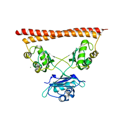
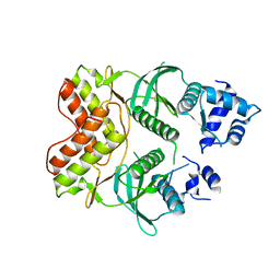
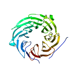
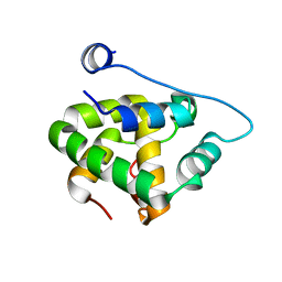
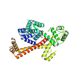
1OF5
 
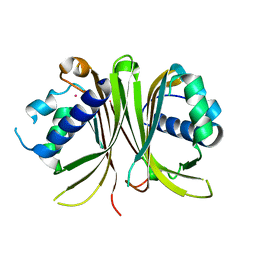 | | Crystal structure of Mex67-Mtr2 | | Descriptor: | MERCURY (II) ION, MRNA EXPORT FACTOR MEX67, MRNA TRANSPORT REGULATOR MTR2 | | Authors: | Fribourg, S, Conti, E. | | Deposit date: | 2003-04-08 | | Release date: | 2003-07-03 | | Last modified: | 2018-06-13 | | Method: | X-RAY DIFFRACTION (2.8 Å) | | Cite: | Structural similarity in the absence of sequence homology of the messenger RNA export factors Mtr2 and p15.
EMBO Rep., 4, 2003
|
|
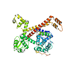
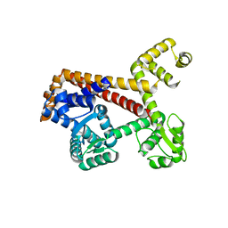
1HL6
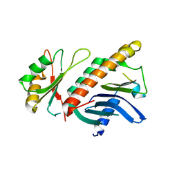 | | A novel mode of RBD-protein recognition in the Y14-mago complex | | Descriptor: | CG8781 PROTEIN, MAGO NASHI PROTEIN | | Authors: | Fribourg, S, Gatfield, D, Yao, W, Izaurralde, E, Conti, E. | | Deposit date: | 2003-03-13 | | Release date: | 2003-05-08 | | Last modified: | 2011-07-13 | | Method: | X-RAY DIFFRACTION (2.5 Å) | | Cite: | A Novel Mode of Rbd-Protein Recognition in the Y14-Mago Complex
Nat.Struct.Biol., 10, 2003
|
|
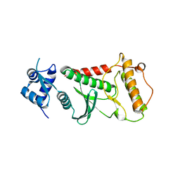
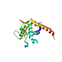
1JKG
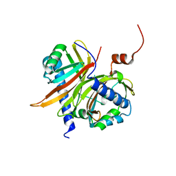 | | Structural basis for the recognition of a nucleoporin FG-repeat by the NTF2-like domain of TAP-p15 mRNA nuclear export factor | | Descriptor: | TAP, p15 | | Authors: | Fribourg, S, Braun, I.C, Izaurralde, E, Conti, E. | | Deposit date: | 2001-07-12 | | Release date: | 2001-10-17 | | Last modified: | 2024-03-13 | | Method: | X-RAY DIFFRACTION (1.9 Å) | | Cite: | Structural basis for the recognition of a nucleoporin FG repeat by the NTF2-like domain of the TAP/p15 mRNA nuclear export factor.
Mol.Cell, 8, 2001
|
|
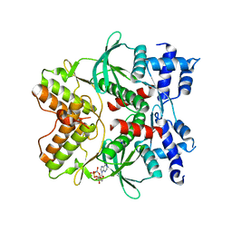
1JN5
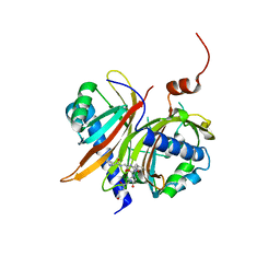 | | Structural basis for the recognition of a nucleoporin FG-repeat by the NTF2-like domain of TAP-p15 mRNA export factor | | Descriptor: | FG-repeat, TAP, p15 | | Authors: | Fribourg, S, Braun, I.C, Izaurralde, E, Conti, E. | | Deposit date: | 2001-07-23 | | Release date: | 2001-10-17 | | Last modified: | 2023-10-25 | | Method: | X-RAY DIFFRACTION (2.8 Å) | | Cite: | Structural basis for the recognition of a nucleoporin FG repeat by the NTF2-like domain of the TAP/p15 mRNA nuclear export factor.
Mol.Cell, 8, 2001
|
|
7PU5
 | | Structure of SFPQ-NONO complex | | Descriptor: | MAGNESIUM ION, Non-POU domain-containing octamer-binding protein, Splicing factor, ... | | Authors: | Fribourg, S. | | Deposit date: | 2021-09-28 | | Release date: | 2022-03-16 | | Last modified: | 2024-01-31 | | Method: | X-RAY DIFFRACTION (2.999 Å) | | Cite: | Crystal structure of SFPQ-NONO heterodimer.
Biochimie, 198, 2022
|
|
5I2T
 | |
5AFQ
 | | Crystal structure of RPC62 - RPC32 beta | | Descriptor: | DNA-DIRECTED RNA POLYMERASE III SUBUNIT RPC3, RPC32 BETA (RPC7L) | | Authors: | Fribourg, S. | | Deposit date: | 2015-01-23 | | Release date: | 2015-10-07 | | Last modified: | 2024-01-10 | | Method: | X-RAY DIFFRACTION (7 Å) | | Cite: | Structural Analysis of Human Rpc32Beta - Rpc62 Complex.
J.Struct.Biol., 192, 2015
|
|
6FDO
 | | Rio2 structure | | Descriptor: | Serine/threonine-protein kinase RIO2 | | Authors: | Fribourg, S. | | Deposit date: | 2017-12-26 | | Release date: | 2019-01-30 | | Last modified: | 2019-10-09 | | Method: | X-RAY DIFFRACTION (2.6 Å) | | Cite: | In vitro dimerization of human RIO2 kinase.
Rna Biol., 16, 2019
|
|
6FDM
 | | Human Rio2 kinase structure | | Descriptor: | PHOSPHOAMINOPHOSPHONIC ACID-ADENYLATE ESTER, SODIUM ION, Serine/threonine-protein kinase RIO2 | | Authors: | Fribourg, S. | | Deposit date: | 2017-12-26 | | Release date: | 2019-01-30 | | Last modified: | 2019-10-09 | | Method: | X-RAY DIFFRACTION (2.1 Å) | | Cite: | In vitro dimerization of human RIO2 kinase.
Rna Biol., 16, 2019
|
|
6FDN
 | | Rio2 structure | | Descriptor: | Serine/threonine-protein kinase RIO2 | | Authors: | Fribourg, S. | | Deposit date: | 2017-12-26 | | Release date: | 2019-01-30 | | Last modified: | 2019-10-09 | | Method: | X-RAY DIFFRACTION (2.9 Å) | | Cite: | In vitro dimerization of human RIO2 kinase.
Rna Biol., 16, 2019
|
|
4C0B
 | | Structure of wild-type Clp1p-Pcf11p (454 -563) complex | | Descriptor: | ADENOSINE-5'-TRIPHOSPHATE, MAGNESIUM ION, MRNA CLEAVAGE AND POLYADENYLATION FACTOR CLP1, ... | | Authors: | Fribourg, S, Dupin, A.F. | | Deposit date: | 2013-08-01 | | Release date: | 2014-02-19 | | Last modified: | 2023-12-20 | | Method: | X-RAY DIFFRACTION (2.77 Å) | | Cite: | Structural basis for ATP loss by Clp1p in a G135R mutant protein.
Biochimie, 101, 2014
|
|
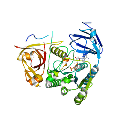
4CLQ
 | | Structure of Rcl1p - Bms1p complex | | Descriptor: | RIBOSOME BIOGENESIS PROTEIN BMS1, RNA 3'-TERMINAL PHOSPHATE CYCLASE-LIKE PROTEIN | | Authors: | Fribourg, S. | | Deposit date: | 2014-01-15 | | Release date: | 2014-11-26 | | Last modified: | 2017-08-23 | | Method: | X-RAY DIFFRACTION (2.02 Å) | | Cite: | Crucial Role of the Rcl1P-Bms1P Interaction for Yeast Pre-Ribosomal RNA Processing.
Nucleic Acids Res., 42, 2014
|
|
5NLG
 | | RRP5 C-terminal domain | | Descriptor: | rRNA biogenesis protein RRP5 | | Authors: | Thore, S, Fribourg, S. | | Deposit date: | 2017-04-04 | | Release date: | 2018-08-29 | | Last modified: | 2018-10-31 | | Method: | X-RAY DIFFRACTION (2.35 Å) | | Cite: | Structural and interaction analysis of the Rrp5 C-terminal region.
FEBS Open Bio, 8, 2018
|
|
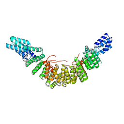
5AMS
 | | Crystal structure of Sqt1 | | Descriptor: | RIBOSOME ASSEMBLY PROTEIN SQT1 | | Authors: | Frenois, F, Legrand, P, Fribourg, S. | | Deposit date: | 2015-08-31 | | Release date: | 2016-01-13 | | Last modified: | 2016-01-20 | | Method: | X-RAY DIFFRACTION (3.35 Å) | | Cite: | Sqt1P is an Eight-Bladed Wd40 Protein
Acta Crystallogr.,Sect.F, 72, 2016
|
|
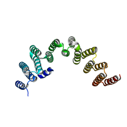
2UY1
 | |
1Z60
 | | Solution structure of the carboxy-terminal domain of human TFIIH P44 subunit | | Descriptor: | TFIIH basal transcription factor complex p44 subunit, ZINC ION | | Authors: | Kellenberger, E, Dominguez, C, Fribourg, S, Wasielewski, E, Moras, D, Poterszman, A, Boelens, R, Kieffer, B. | | Deposit date: | 2005-03-21 | | Release date: | 2005-04-12 | | Last modified: | 2021-11-10 | | Method: | SOLUTION NMR | | Cite: | Solution structure of the C-terminal domain of TFIIH P44 subunit reveals a novel type of C4C4 ring domain involved in protein-protein interactions.
J.Biol.Chem., 280, 2005
|
|
2JLT
 | | Crystal structure of an RNA kissing complex | | Descriptor: | R06, TAR | | Authors: | DiPrimo, C, Fribourg, S. | | Deposit date: | 2008-09-15 | | Release date: | 2009-08-18 | | Last modified: | 2017-07-12 | | Method: | X-RAY DIFFRACTION (2.9 Å) | | Cite: | Exploring Tar-RNA Aptamer Loop-Loop Interaction by X-Ray Crystallography, Uv Spectroscopy and Surface Plasmon Resonance.
Nucleic Acids Res., 36, 2008
|
|
2L9B
 | | Heterodimer between Rna14p monkeytail domain and Rna15p hinge domain of the yeast CF IA complex | | Descriptor: | mRNA 3'-end-processing protein RNA14, mRNA 3'-end-processing protein RNA15 | | Authors: | Moreno-Morcillo, M, Minvielle-Sebastia, L, Fribourg, S, Mackereth, C.D. | | Deposit date: | 2011-02-07 | | Release date: | 2011-04-27 | | Last modified: | 2023-06-14 | | Method: | SOLUTION NMR | | Cite: | Locked Tether Formation by Cooperative Folding of Rna14p Monkeytail and Rna15p Hinge Domains in the Yeast CF IA Complex.
Structure, 19, 2011
|
|
4C0H
 | |
2BR2
 | | RNase PH core of the archaeal exosome | | Descriptor: | CHLORIDE ION, EXOSOME COMPLEX EXONUCLEASE 1, EXOSOME COMPLEX EXONUCLEASE 2 | | Authors: | lorentzen, E, Fribourg, S, Conti, E. | | Deposit date: | 2005-04-30 | | Release date: | 2005-06-06 | | Last modified: | 2023-12-13 | | Method: | X-RAY DIFFRACTION (2.8 Å) | | Cite: | The Archaeal Exosome Core is a Hexameric Ring Structure with Three Catalytic Subunits.
Nat.Struct.Mol.Biol., 12, 2005
|
|
6HFA
 | | Crystal structure of hDM2 in complex with a C-terminal triurea capped peptide chimera foldamer. | | Descriptor: | E3 ubiquitin-protein ligase Mdm2, LM266, 1-[(2~{S})-2-azanyl-3-methyl-butyl]urea | | Authors: | Buratto, J, Mauran, L, Goudreau, S, Fribourg, S, Guichard, G. | | Deposit date: | 2018-08-21 | | Release date: | 2020-07-08 | | Last modified: | 2023-11-15 | | Method: | X-RAY DIFFRACTION (1.79 Å) | | Cite: | Structural Basis for alpha-Helix Mimicry and Inhibition of Protein-Protein Interactions with Oligourea Foldamers.
Angew.Chem.Int.Ed.Engl., 2020
|
|
2XV4
 | | Structure of Human RPC62 (partial) | | Descriptor: | DNA-DIRECTED RNA POLYMERASE III SUBUNIT RPC3, PHOSPHATE ION | | Authors: | Lefevre, S, Legrand, P, Fribourg, S. | | Deposit date: | 2010-10-22 | | Release date: | 2011-03-02 | | Last modified: | 2011-07-13 | | Method: | X-RAY DIFFRACTION (2.95 Å) | | Cite: | Structure-Function Analysis of Hrpc62 Provides Insights Into RNA Polymerase III Transcription
Nat.Struct.Mol.Biol., 18, 2011
|
|
2XUB
 | | Human RPC62 subunit structure | | Descriptor: | DNA-DIRECTED RNA POLYMERASE III SUBUNIT RPC3 | | Authors: | Lefevre, S, Legrand, P, Fribourg, S. | | Deposit date: | 2010-10-18 | | Release date: | 2011-03-02 | | Last modified: | 2011-07-13 | | Method: | X-RAY DIFFRACTION (2.8 Å) | | Cite: | Structure-Function Analysis of Hrpc62 Provides Insights Into RNA Polymerase III Transcription
Nat.Struct.Mol.Biol., 18, 2011
|
|
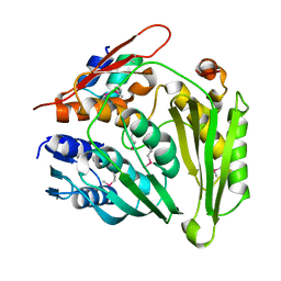
2V7F
 | | Structure of P. abyssi RPS19 protein | | Descriptor: | CHLORIDE ION, RPS19E SSU RIBOSOMAL PROTEIN S19E | | Authors: | Gregory, L.A, Aguissa-Toure, A.H, Pinaud, N, Legrand, P, Gleizes, P.E, Fribourg, S. | | Deposit date: | 2007-07-30 | | Release date: | 2007-09-11 | | Last modified: | 2011-07-13 | | Method: | X-RAY DIFFRACTION (1.15 Å) | | Cite: | Molecular Basis of Diamond Blackfan Anemia: Structure and Function Analysis of Rps19.
Nucleic Acids Res., 35, 2007
|
|
2V94
 | |


