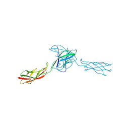
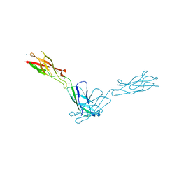
Loading
PDBj
MenuPDBj@FacebookPDBj@TwitterPDBj@YouTubewwPDB FoundationwwPDB
RCSB PDBPDBeBMRBAdv. SearchSearch help
Search by PDB author
2QVF
DownloadVisualize
BU of 2qvf by Molmil
mouse E-cadherin domains 1,2
Descriptor: CALCIUM ION, Epithelial-cadherin (E-cadherin) (Uvomorulin) (Cadherin-1) (ARC-1) (CD324 antigen)
Authors:Carroll, K.J.
Deposit date:2007-08-08
Release date:2008-08-12
Last modified:2023-08-30
Method:X-RAY DIFFRACTION (2.4 Å)
Cite:E-cadherin domains 1,2
To be Published
1S31
DownloadVisualize
BU of 1s31 by Molmil
Crystal Structure Analysis of the human Tub protein (isoform a) spanning residues 289 through 561
Descriptor: TRIETHYLENE GLYCOL, tubby isoform a
Authors:Boutboul, S, Carroll, K.J, Basdevant, A, Gomez, C, Nandrot, E, Clement, K, Shapiro, L, Abitbol, M.
Deposit date:2004-01-12
Release date:2005-01-25
Last modified:2023-08-23
Method:X-RAY DIFFRACTION (2.704 Å)
Cite:A novel human obesity and sensory deficit syndrome resulting from a mutation in the TUB gene
To be Published
2QVI
DownloadVisualize
BU of 2qvi by Molmil
Crystal structure of N-cadherin domains EC12
Descriptor: CALCIUM ION, Cadherin-2
Authors:Shapiro, L.S, Carroll, K.J.
Deposit date:2007-08-08
Release date:2008-08-12
Last modified:2024-02-21
Method:X-RAY DIFFRACTION (3.012 Å)
Cite:Biophysical characterization of N-cadherin and E-cadherin homophilic and heterophilic interactions
To be Published
3LNG
DownloadVisualize
BU of 3lng by Molmil
Crystal structure of E-cadherin EC12 AA extension
Descriptor: CALCIUM ION, Cadherin-1
Authors:Harrison, O, Jin, X, Shapiro, L.
Deposit date:2010-02-02
Release date:2010-03-02
Last modified:2023-09-06
Method:X-RAY DIFFRACTION (2.7 Å)
Cite:Two-step adhesive binding by classical cadherins.
Nat.Struct.Mol.Biol., 17, 2010
3LND
DownloadVisualize
BU of 3lnd by Molmil
Crystal structure of cadherin-6 EC12 W4A
Descriptor: CALCIUM ION, Cdh6 protein
Authors:Jin, X, Harrison, O, Shapiro, L.
Deposit date:2010-02-02
Release date:2010-03-02
Last modified:2024-02-21
Method:X-RAY DIFFRACTION (2.82 Å)
Cite:Two-step adhesive binding by classical cadherins.
Nat.Struct.Mol.Biol., 17, 2010
3LNE
DownloadVisualize
BU of 3lne by Molmil
Crystal structure of E-cadherin EC12 K14E
Descriptor: CALCIUM ION, Cadherin-1, GLYCEROL
Authors:Jin, X, Harrison, O, Shapiro, L.
Deposit date:2010-02-02
Release date:2010-03-02
Last modified:2023-09-06
Method:X-RAY DIFFRACTION (2 Å)
Cite:Two-step adhesive binding by classical cadherins.
Nat.Struct.Mol.Biol., 17, 2010
3LNF
DownloadVisualize
BU of 3lnf by Molmil
Crystal structure of E-cadherin EC12 K14EW2A
Descriptor: CALCIUM ION, Cadherin-1
Authors:Jin, X, Harrison, O, Shapiro, L.
Deposit date:2010-02-02
Release date:2010-03-02
Last modified:2023-09-06
Method:X-RAY DIFFRACTION (2.5 Å)
Cite:Two-step adhesive binding by classical cadherins.
Nat.Struct.Mol.Biol., 17, 2010
3LNI
DownloadVisualize
BU of 3lni by Molmil
Crystal structure of E-cadherin EC12 E89A
Descriptor: CALCIUM ION, Cadherin-1
Authors:Harrison, O, Jin, X, Shapiro, L.
Deposit date:2010-02-02
Release date:2010-03-02
Last modified:2023-09-06
Method:X-RAY DIFFRACTION (2.3 Å)
Cite:Two-step adhesive binding by classical cadherins.
Nat.Struct.Mol.Biol., 17, 2010
3LNH
DownloadVisualize
BU of 3lnh by Molmil
Crystal structure of E-cadherin EC12 W2A
Descriptor: CALCIUM ION, Cadherin-1
Authors:Harrison, O, Jin, X, Shapiro, L.
Deposit date:2010-02-02
Release date:2010-03-02
Last modified:2023-09-06
Method:X-RAY DIFFRACTION (2.6 Å)
Cite:Two-step adhesive binding by classical cadherins.
Nat.Struct.Mol.Biol., 17, 2010

218853

PDB entries from 2024-04-24

PDB statisticsPDBj update infoContact PDBjnumon