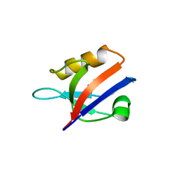
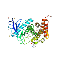
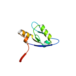
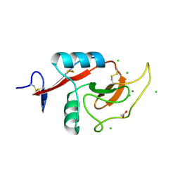
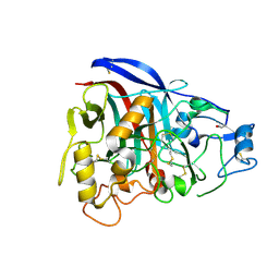
4R2Z
 
 | |
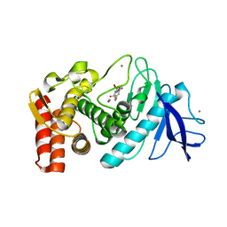
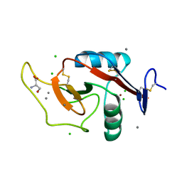
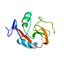
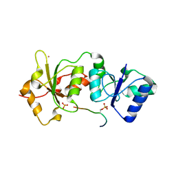
4LG0
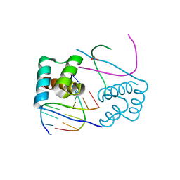
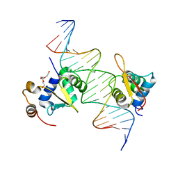 | | Structure of a ternary FOXO1-ETS1 DNA complex | | Descriptor: | CALCIUM ION, DNA (5'-D(*DAP*DAP*DAP*DCP*DAP*DAP*DTP*DAP*DAP*DCP*DAP*DGP*DGP*DAP*DAP*DAP*DCP*DCP*DGP*DTP*DG)-3'), DNA (5'-D(*DTP*DTP*DCP*DAP*DCP*DGP*DGP*DTP*DTP*DTP*DCP*DCP*DTP*DGP*DTP*DTP*DAP*DTP*DTP*DGP*DT)-3'), ... | | Authors: | Birrane, G, Choy, W.C, Datta, D, Geiger, C.A, Grant, M.A. | | Deposit date: | 2013-06-27 | | Release date: | 2014-07-02 | | Last modified: | 2024-02-28 | | Method: | X-RAY DIFFRACTION (2.19 Å) | | Cite: | Structure of a ternary FOXO1-ETS1 DNA complex
To be Published
|
|
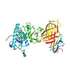
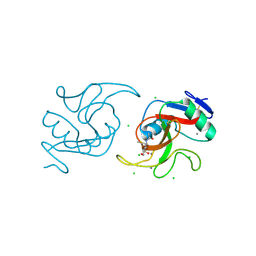
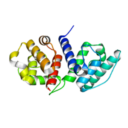
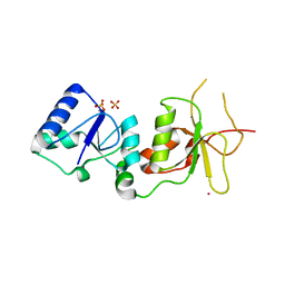
6E7K
 | |
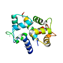
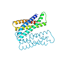
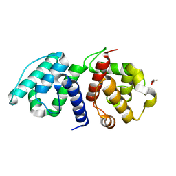
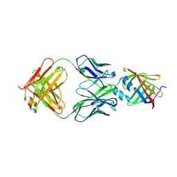
3BYA
 | |
3CMY
 | | Structure of a homeodomain in complex with DNA | | Descriptor: | 1,2-ETHANEDIOL, 5'-D(*DAP*DCP*DAP*DTP*DAP*DAP*DP*DCP*DGP*DAP*DTP*DTP*DAP*DC)-3', 5'-D(*DTP*DGP*DTP*DAP*DAP*DTP*DCP*DGP*DAP*DTP*DTP*DAP*DTP*DG)-3', ... | | Authors: | Birrane, G, Ladias, J.A.A, Soni, A. | | Deposit date: | 2008-03-24 | | Release date: | 2009-02-17 | | Last modified: | 2024-04-03 | | Method: | X-RAY DIFFRACTION (1.95 Å) | | Cite: | Structural Basis for DNA Recognition by the Human PAX3 Homeodomain.
Biochemistry, 48, 2009
|
|
2NTE
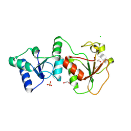 | | Crystal Structure of the BARD1 BRCT Domains | | Descriptor: | 1,2-ETHANEDIOL, BRCA1-associated RING domain protein 1, CHLORIDE ION, ... | | Authors: | Birrane, G, Varma, A.K, Soni, A, Ladias, J.A.A. | | Deposit date: | 2006-11-07 | | Release date: | 2007-06-12 | | Last modified: | 2023-12-27 | | Method: | X-RAY DIFFRACTION (1.9 Å) | | Cite: | Crystal structure of the BARD1 BRCT domains.
Biochemistry, 46, 2007
|
|
4XWH
 | | Crystal structure of the human N-acetyl-alpha-glucosaminidase | | Descriptor: | 2-acetamido-2-deoxy-beta-D-glucopyranose, 2-acetamido-2-deoxy-beta-D-glucopyranose-(1-4)-2-acetamido-2-deoxy-beta-D-glucopyranose, Alpha-N-acetylglucosaminidase, ... | | Authors: | Birrane, G, Meiyappan, M, Dassier, A. | | Deposit date: | 2015-01-28 | | Release date: | 2016-02-03 | | Last modified: | 2023-09-27 | | Method: | X-RAY DIFFRACTION (2.32 Å) | | Cite: | Structural characterization of the alpha-N-acetylglucosaminidase, a key enzyme in the pathogenesis of Sanfilippo syndrome B.
J.Struct.Biol., 205, 2019
|
|
1MFG
 | |
1MFL
 | |
3QH5
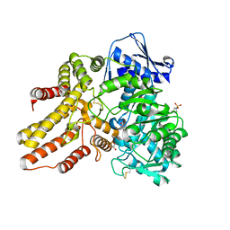
 | | Structure of Thermolysin in complex with N-Carbobenzyloxy-L-aspartic acid and L-Phenylalanine Methyl Ester | | Descriptor: | CALCIUM ION, DI(HYDROXYETHYL)ETHER, N-[(benzyloxy)carbonyl]-L-aspartic acid, ... | | Authors: | Birrane, G, Bhyravbhatla, B, Navia, M. | | Deposit date: | 2011-01-25 | | Release date: | 2012-01-04 | | Last modified: | 2023-12-06 | | Method: | X-RAY DIFFRACTION (1.5 Å) | | Cite: | Synthesis of Aspartame by Thermolysin: An X-ray Structural Study.
ACS MED.CHEM.LETT., 5, 2014
|
|
3QGO
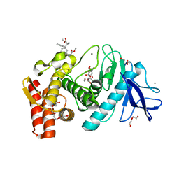
 | | Structure of Thermolysin in complex with L-Phenylalanine methylester | | Descriptor: | ACETATE ION, CALCIUM ION, DIMETHYL SULFOXIDE, ... | | Authors: | Birrane, G, Bhyravbhatla, B, Navia, M. | | Deposit date: | 2011-01-24 | | Release date: | 2012-01-04 | | Last modified: | 2023-12-06 | | Method: | X-RAY DIFFRACTION (1.45 Å) | | Cite: | Synthesis of Aspartame by Thermolysin: An X-ray Structural Study.
ACS MED.CHEM.LETT., 5, 2014
|
|
3QH1
 | | Structure of Thermolysin in complex with N-benzyloxycarbonyl-L-aspartic acid | | Descriptor: | CALCIUM ION, N-[(benzyloxy)carbonyl]-L-aspartic acid, Thermolysin, ... | | Authors: | Birrane, G, Bhyravbhatla, B, Navia, M. | | Deposit date: | 2011-01-25 | | Release date: | 2012-01-04 | | Last modified: | 2023-09-13 | | Method: | X-RAY DIFFRACTION (1.55 Å) | | Cite: | Synthesis of Aspartame by Thermolysin: An X-ray Structural Study.
ACS MED.CHEM.LETT., 5, 2014
|
|
6XIY
 | | Crystal Structure of the Carbohydrate Recognition Domain of the Human Macrophage Galactose C-Type Lectin Bound to methyl 2-(acetylamino)-2-deoxy-1-thio-alpha-D-galactopyranose | | Descriptor: | C-type lectin domain family 10 member A, CALCIUM ION, CHLORIDE ION, ... | | Authors: | Birrane, G, Murphy, P.V, Gabba, A, Luz, J.G. | | Deposit date: | 2020-06-22 | | Release date: | 2021-03-31 | | Last modified: | 2023-10-18 | | Method: | X-RAY DIFFRACTION (2.307 Å) | | Cite: | Crystal Structure of the Carbohydrate Recognition Domain of the Human Macrophage Galactose C-Type Lectin Bound to GalNAc and the Tumor-Associated Tn Antigen.
Biochemistry, 60, 2021
|
|
6N63
 | | Crystal structure of an Iron binding protein | | Descriptor: | ACETATE ION, ENCAPSULIN CARGO PROTEIN, FE (III) ION, ... | | Authors: | Birrane, G, Geissen, T.W. | | Deposit date: | 2018-11-24 | | Release date: | 2019-07-17 | | Last modified: | 2024-04-03 | | Method: | X-RAY DIFFRACTION (1.72 Å) | | Cite: | Large protein organelles form a new iron sequestration system with high storage capacity.
Elife, 8, 2019
|
|
4F8K
 | | Molecular analysis of the interaction between the prostacyclin receptor and the first PDZ domain of PDZK1 | | Descriptor: | Na(+)/H(+) exchange regulatory cofactor NHE-RF3, Prostacyclin receptor | | Authors: | Kocher, O, Birrane, G, Kinsella, B.T, Mulvaney, E.P. | | Deposit date: | 2012-05-17 | | Release date: | 2013-02-27 | | Last modified: | 2023-09-13 | | Method: | X-RAY DIFFRACTION (1.7 Å) | | Cite: | Molecular Analysis of the Prostacyclin Receptor's Interaction with the PDZ1 Domain of Its Adaptor Protein PDZK1.
Plos One, 8, 2013
|
|
6W12
 | | Crystal Structure of the Carbohydrate Recognition Domain of the Human Macrophage Galactose C-Type Lectin Bound to the Tumor-Associated Tn Antigen | | Descriptor: | 2-acetamido-2-deoxy-alpha-D-galactopyranose, C-type lectin domain family 10 member A, CALCIUM ION, ... | | Authors: | Birrane, G, Murphy, P.V, Gabba, A, Luz, J.G. | | Deposit date: | 2020-03-03 | | Release date: | 2021-03-10 | | Last modified: | 2023-10-18 | | Method: | X-RAY DIFFRACTION (2 Å) | | Cite: | Crystal Structure of the Carbohydrate Recognition Domain of the Human Macrophage Galactose C-Type Lectin Bound to GalNAc and the Tumor-Associated Tn Antigen.
Biochemistry, 60, 2021
|
|
6O31
 | |
6OA6
 | |
6PY1
 | | Crystal Structure of the Carbohydrate Recognition Domain of the Human Macrophage Galactose C-Type Lectin Bound to GalNAc | | Descriptor: | 2-acetamido-2-deoxy-alpha-D-galactopyranose, ACETATE ION, C-type lectin domain family 10 member A, ... | | Authors: | Birrane, G, Murphy, P.V, Gabba, A, Luz, J.G. | | Deposit date: | 2019-07-28 | | Release date: | 2020-07-29 | | Last modified: | 2023-10-11 | | Method: | X-RAY DIFFRACTION (1.701 Å) | | Cite: | Crystal Structure of the Carbohydrate Recognition Domain of the Human Macrophage Galactose C-Type Lectin Bound to GalNAc and the Tumor-Associated Tn Antigen.
Biochemistry, 60, 2021
|
|
6PUV
 | | Crystal Structure of the Carbohydrate Recognition Domain of the Human Macrophage Galactose C-Type Lectin | | Descriptor: | C-type lectin domain family 10 member A, CALCIUM ION | | Authors: | Birrane, G, Murphy, P.V, Gabba, A, Luz, J.G. | | Deposit date: | 2019-07-18 | | Release date: | 2020-07-22 | | Last modified: | 2023-10-11 | | Method: | X-RAY DIFFRACTION (1.2 Å) | | Cite: | Crystal Structure of the Carbohydrate Recognition Domain of the Human Macrophage Galactose C-Type Lectin Bound to GalNAc and the Tumor-Associated Tn Antigen.
Biochemistry, 60, 2021
|
|
2ING
 | | X-ray Structure of the BRCA1 BRCT mutant M1775K | | Descriptor: | Breast cancer type 1 susceptibility protein, COBALT (II) ION, SULFATE ION | | Authors: | Birrane, G, Soni, A, Ladias, J.A.A. | | Deposit date: | 2006-10-07 | | Release date: | 2007-09-04 | | Last modified: | 2023-08-30 | | Method: | X-RAY DIFFRACTION (3.6 Å) | | Cite: | Pathogenicity of the BRCA1 missense variant M1775K is determined by the disruption of the BRCT phosphopeptide-binding pocket: a multi-modal approach.
Eur.J.Hum.Genet., 16, 2008
|
|
5D8J
 | |
1Q9H
 | | 3-Dimensional structure of native Cel7A from Talaromyces emersonii | | Descriptor: | 2-acetamido-2-deoxy-beta-D-glucopyranose, 2-acetamido-2-deoxy-beta-D-glucopyranose-(1-4)-2-acetamido-2-deoxy-beta-D-glucopyranose, cellobiohydrolase I catalytic domain | | Authors: | Grassick, A, Thompson, R, Murray, P.G, Collins, C.M, Byrnes, L, Tuohy, M.G, Birrane, G, Higgins, T.M. | | Deposit date: | 2003-08-25 | | Release date: | 2004-11-09 | | Last modified: | 2020-07-29 | | Method: | X-RAY DIFFRACTION (2.35 Å) | | Cite: | Three-dimensional structure of a thermostable native cellobiohydrolase, CBH IB, and molecular characterization of the cel7 gene from the filamentous fungus, Talaromyces emersonii
Eur.J.Biochem., 271, 2004
|
|
1Y98
 | | Structure of the BRCT repeats of BRCA1 bound to a CtIP phosphopeptide. | | Descriptor: | Breast cancer type 1 susceptibility protein, COBALT (II) ION, CtIP PHOSPHORYLATED PEPTIDE, ... | | Authors: | Varma, A.K, Brown, R.S, Birrane, G, Ladias, J.A.A. | | Deposit date: | 2004-12-14 | | Release date: | 2005-08-30 | | Last modified: | 2023-08-23 | | Method: | X-RAY DIFFRACTION (2.5 Å) | | Cite: | Structural Basis for Cell Cycle Checkpoint Control by the BRCA1-CtIP Complex.
Biochemistry, 44, 2005
|
|
2OEI
 | |


