Loading
PDBj
MenuPDBj@FacebookPDBj@TwitterPDBj@YouTubewwPDB FoundationwwPDB
RCSB PDBPDBeBMRBAdv. SearchSearch help
Search by PDB author
1BP1
DownloadVisualize
BU of 1bp1 by Molmil
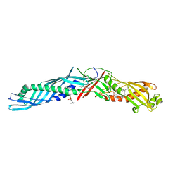
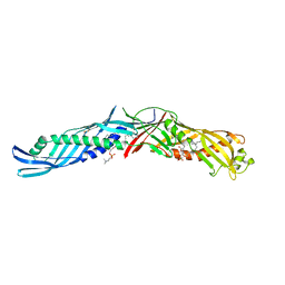
CRYSTAL STRUCTURE OF BPI, THE HUMAN BACTERICIDAL PERMEABILITY-INCREASING PROTEIN
Descriptor: 1,2-DIACYL-SN-GLYCERO-3-PHOSPHOCHOLINE, BACTERICIDAL/PERMEABILITY-INCREASING PROTEIN
Authors:Beamer, L.J, Carroll, S.F, Eisenberg, D.
Deposit date:1997-04-08
Release date:1997-09-04
Last modified:2021-11-03
Method:X-RAY DIFFRACTION (2.4 Å)
Cite:Crystal structure of human BPI and two bound phospholipids at 2.4 angstrom resolution.
Science, 276, 1997
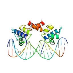
1LMB
DownloadVisualize
BU of 1lmb by Molmil
REFINED 1.8 ANGSTROM CRYSTAL STRUCTURE OF THE LAMBDA REPRESSOR-OPERATOR COMPLEX
Descriptor: DNA (5'-D(*AP*AP*TP*AP*CP*CP*AP*CP*TP*GP*GP*CP*GP*GP*TP*GP*A P*TP*AP*T)-3'), DNA (5'-D(*TP*AP*TP*AP*TP*CP*AP*CP*CP*GP*CP*CP*AP*GP*TP*GP*G P*TP*AP*T)-3'), PROTEIN (LAMBDA REPRESSOR)
Authors:Beamer, L.J, Pabo, C.O.
Deposit date:1991-11-05
Release date:1991-11-05
Last modified:2024-02-14
Method:X-RAY DIFFRACTION (1.8 Å)
Cite:Refined 1.8 A crystal structure of the lambda repressor-operator complex.
J.Mol.Biol., 227, 1992
1ZGK
DownloadVisualize
BU of 1zgk by Molmil
1.35 angstrom structure of the Kelch domain of Keap1
Descriptor: Kelch-like ECH-associated protein 1
Authors:Li, X, Bottoms, C.A, Hannink, M, Beamer, L.J.
Deposit date:2005-04-21
Release date:2005-10-04
Last modified:2011-07-13
Method:X-RAY DIFFRACTION (1.35 Å)
Cite:Conserved solvent and side-chain interactions in the 1.35 Angstrom structure of the Kelch domain of Keap1.
Acta Crystallogr.,Sect.D, 61, 2005
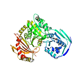
5JN5
DownloadVisualize
BU of 5jn5 by Molmil
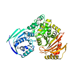
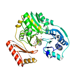
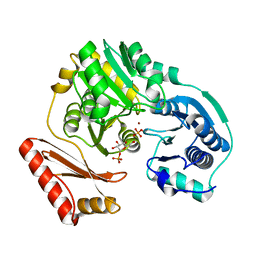
Crystal structure of the D263Y missense variant of human PGM1
Descriptor: CALCIUM ION, Phosphoglucomutase-1, SULFATE ION
Authors:Beamer, L.J, Stiers, K.M.
Deposit date:2016-04-29
Release date:2017-02-08
Last modified:2019-11-27
Method:X-RAY DIFFRACTION (1.75 Å)
Cite:Asp263 missense variants perturb the active site of human phosphoglucomutase 1.
FEBS J., 284, 2017
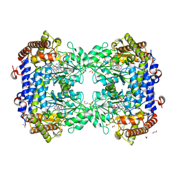
8FSD
DownloadVisualize
BU of 8fsd by Molmil
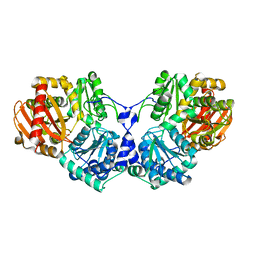
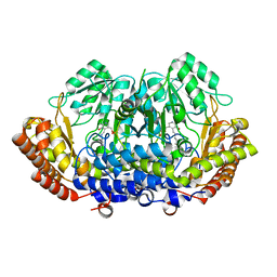
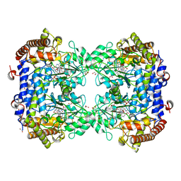
P130R mutant of soybean SHMT8 in complex with PLP-glycine and formylTHF
Descriptor: 1,2-ETHANEDIOL, N-GLYCINE-[3-HYDROXY-2-METHYL-5-PHOSPHONOOXYMETHYL-PYRIDIN-4-YL-METHANE], N-[4-({[(6S)-2-amino-5-formyl-4-oxo-3,4,5,6,7,8-hexahydropteridin-6-yl]methyl}amino)benzoyl]-L-glutamic acid, ...
Authors:Beamer, L.J, Korasick, D.A.
Deposit date:2023-01-09
Release date:2023-10-18
Last modified:2024-01-31
Method:X-RAY DIFFRACTION (1.49 Å)
Cite:Structural and functional analysis of two SHMT8 variants associated with soybean cyst nematode resistance.
Febs J., 291, 2024
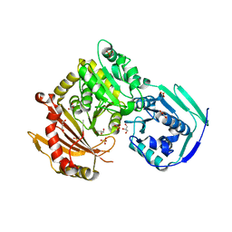
5EPC
DownloadVisualize
BU of 5epc by Molmil
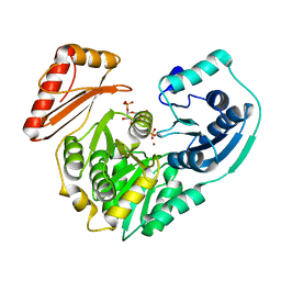
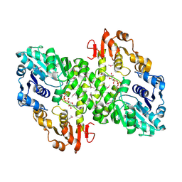
Crystal structure of wild-type human phosphoglucomutase 1
Descriptor: GLYCEROL, MAGNESIUM ION, Phosphoglucomutase-1, ...
Authors:Beamer, L.J.
Deposit date:2015-11-11
Release date:2016-04-27
Last modified:2023-09-27
Method:X-RAY DIFFRACTION (1.85 Å)
Cite:Induced Structural Disorder as a Molecular Mechanism for Enzyme Dysfunction in Phosphoglucomutase 1 Deficiency.
J.Mol.Biol., 428, 2016
5F9C
DownloadVisualize
BU of 5f9c by Molmil
Crystal structure of the G121R mutant of human phosphoglucomutase 1
Descriptor: GLYCEROL, MAGNESIUM ION, Phosphoglucomutase-1, ...
Authors:Beamer, L.J.
Deposit date:2015-12-09
Release date:2016-04-27
Last modified:2023-09-27
Method:X-RAY DIFFRACTION (2.5 Å)
Cite:Induced Structural Disorder as a Molecular Mechanism for Enzyme Dysfunction in Phosphoglucomutase 1 Deficiency.
J.Mol.Biol., 428, 2016
2H4L
DownloadVisualize
BU of 2h4l by Molmil
Complex of PMM/PGM with ribose 1-phosphate
Descriptor: 1-O-phosphono-alpha-D-ribofuranose, Phosphomannomutase/phosphoglucomutase, ZINC ION
Authors:Beamer, L.J.
Deposit date:2006-05-24
Release date:2006-08-08
Last modified:2023-08-30
Method:X-RAY DIFFRACTION (2.4 Å)
Cite:Complexes of the enzyme phosphomannomutase/phosphoglucomutase with a slow substrate and an inhibitor.
Acta Crystallogr.,Sect.F, 62, 2006
6N1E
DownloadVisualize
BU of 6n1e by Molmil
Crystal structure of X. citri phosphoglucomutase in complex with 1-methyl-glucose 6-phosphate
Descriptor: 1-deoxy-7-O-phosphono-alpha-D-gluco-hept-2-ulopyranose, MAGNESIUM ION, Phosphomannomutase/phosphoglucomutase
Authors:Beamer, L.J, Stiers, K.M.
Deposit date:2018-11-08
Release date:2019-05-01
Last modified:2023-10-11
Method:X-RAY DIFFRACTION (1.7 Å)
Cite:Synthesis, Derivatization, and Structural Analysis of Phosphorylated Mono-, Di-, and Trifluorinated d-Gluco-heptuloses by Glucokinase: Tunable Phosphoglucomutase Inhibition.
Acs Omega, 4, 2019
6UO6
DownloadVisualize
BU of 6uo6 by Molmil
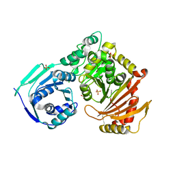
Crystal Structure of the R422Q missense variant of human PGM1
Descriptor: MAGNESIUM ION, Phosphoglucomutase-1, SULFATE ION
Authors:Beamer, L.J, Stiers, K.M.
Deposit date:2019-10-14
Release date:2020-03-04
Last modified:2023-10-11
Method:X-RAY DIFFRACTION (2.15 Å)
Cite:A missense variant remote from the active site impairs stability of human phosphoglucomutase 1.
J. Inherit. Metab. Dis., 43, 2020
5TR2
DownloadVisualize
BU of 5tr2 by Molmil
Crystal structure of the D263G missense variant of human PGM1
Descriptor: CALCIUM ION, GLYCEROL, Phosphoglucomutase-1, ...
Authors:Beamer, L.J, Stiers, K.M.
Deposit date:2016-10-25
Release date:2017-02-08
Last modified:2023-10-04
Method:X-RAY DIFFRACTION (2.5 Å)
Cite:Asp263 missense variants perturb the active site of human phosphoglucomutase 1.
FEBS J., 284, 2017
3OLP
DownloadVisualize
BU of 3olp by Molmil
Crystal structure of a bacterial phosphoglucomutase, an enzyme important in the virulence of multiple human pathogens
Descriptor: MAGNESIUM ION, PHOSPHATE ION, Phosphoglucomutase
Authors:Beamer, L.J, Mehra-Chaudhary, R.
Deposit date:2010-08-26
Release date:2011-02-16
Last modified:2023-09-06
Method:X-RAY DIFFRACTION (1.95 Å)
Cite:Crystal structure of a bacterial phosphoglucomutase, an enzyme involved in the virulence of multiple human pathogens.
Proteins, 79, 2011
1EWF
DownloadVisualize
BU of 1ewf by Molmil
THE 1.7 ANGSTROM CRYSTAL STRUCTURE OF BPI
Descriptor: 1,2-DIACYL-SN-GLYCERO-3-PHOSPHOCHOLINE, BACTERICIDAL/PERMEABILITY-INCREASING PROTEIN
Authors:Kleiger, G, Beamer, L.J, Grothe, R, Mallick, P, Eisenberg, D.
Deposit date:2000-04-25
Release date:2000-06-21
Last modified:2021-11-03
Method:X-RAY DIFFRACTION (1.7 Å)
Cite:The 1.7 A crystal structure of BPI: a study of how two dissimilar amino acid sequences can adopt the same fold.
J.Mol.Biol., 299, 2000
1U6D
DownloadVisualize
BU of 1u6d by Molmil
Crystal structure of the Kelch domain of human Keap1
Descriptor: kelch-like ECH-associated protein 1
Authors:Li, X, Zhang, D, Hannink, M, Beamer, L.J.
Deposit date:2004-07-29
Release date:2004-10-12
Last modified:2024-02-14
Method:X-RAY DIFFRACTION (1.85 Å)
Cite:Crystal structure of the kelch domain of human keap1
J.Biol.Chem., 279, 2004
5HSH
DownloadVisualize
BU of 5hsh by Molmil
Crystal structure of the G291R mutant of human phosphoglucomutase 1
Descriptor: Phosphoglucomutase-1, SULFATE ION
Authors:Stiers, K.M, Beamer, L.J.
Deposit date:2016-01-25
Release date:2016-04-27
Last modified:2023-09-27
Method:X-RAY DIFFRACTION (2.65 Å)
Cite:Induced Structural Disorder as a Molecular Mechanism for Enzyme Dysfunction in Phosphoglucomutase 1 Deficiency.
J.Mol.Biol., 428, 2016
8DOM
DownloadVisualize
BU of 8dom by Molmil
Structure of the N358Y single variant ofserine hydroxymethyltransferase 8 from Glycine max cultivar Essex complexed with PLP
Descriptor: 1,2-ETHANEDIOL, Serine hydroxymethyltransferase
Authors:Korasick, D.A, Beamer, L.J.
Deposit date:2022-07-13
Release date:2023-07-19
Last modified:2024-01-31
Method:X-RAY DIFFRACTION (1.89 Å)
Cite:Structural and functional analysis of two SHMT8 variants associated with soybean cyst nematode resistance.
Febs J., 291, 2024
8DSK
DownloadVisualize
BU of 8dsk by Molmil
Structure of the N358Y variant of serine hydroxymethyltransferase 8 in complex with PLP, glycine, and formyl tetrahydrofolate
Descriptor: 1,2-ETHANEDIOL, N-GLYCINE-[3-HYDROXY-2-METHYL-5-PHOSPHONOOXYMETHYL-PYRIDIN-4-YL-METHANE], N-[4-({[(6S)-2-amino-5-formyl-4-oxo-3,4,5,6,7,8-hexahydropteridin-6-yl]methyl}amino)benzoyl]-L-glutamic acid, ...
Authors:Korasick, D.A, Beamer, L.J.
Deposit date:2022-07-22
Release date:2023-10-18
Last modified:2024-01-31
Method:X-RAY DIFFRACTION (1.63 Å)
Cite:Structural and functional analysis of two SHMT8 variants associated with soybean cyst nematode resistance.
Febs J., 291, 2024
2H5A
DownloadVisualize
BU of 2h5a by Molmil
Complex of the enzyme PMM/PGM with xylose 1-phosphate
Descriptor: 1-O-phosphono-alpha-D-xylopyranose, Phosphomannomutase/phosphoglucomutase, ZINC ION
Authors:Regni, C, Shackelford, G.S, Beamer, L.J.
Deposit date:2006-05-25
Release date:2006-08-08
Last modified:2020-07-29
Method:X-RAY DIFFRACTION (1.72 Å)
Cite:Complexes of the enzyme phosphomannomutase/phosphoglucomutase with a slow substrate and an inhibitor.
Acta Crystallogr.,Sect.F, 62, 2006
2FKF
DownloadVisualize
BU of 2fkf by Molmil
Phosphomannomutase/Phosphoglucomutase from Pseudomonas aeruginosa with alpha-D-glucose 1,6-bisphosphate bound
Descriptor: 1,6-di-O-phosphono-alpha-D-glucopyranose, Phosphomannomutase/phosphoglucomutase, ZINC ION
Authors:Regni, C.A, Beamer, L.J.
Deposit date:2006-01-04
Release date:2006-04-04
Last modified:2023-08-30
Method:X-RAY DIFFRACTION (2 Å)
Cite:The reaction of phosphohexomutase from Pseudomonas aeruginosa: structural insights into a simple processive enzyme.
J.Biol.Chem., 281, 2006
2FKM
DownloadVisualize
BU of 2fkm by Molmil
PMM/PGM S108D mutant with alpha-d-glucose 1,6-bisphosphate bound
Descriptor: 1,6-di-O-phosphono-alpha-D-glucopyranose, Phosphomannomutase/phosphoglucomutase, ZINC ION
Authors:Regni, C.A, Beamer, L.J.
Deposit date:2006-01-04
Release date:2006-04-04
Last modified:2023-08-30
Method:X-RAY DIFFRACTION (1.9 Å)
Cite:The reaction of phosphohexomutase from Pseudomonas aeruginosa: structural insights into a simple processive enzyme.
J.Biol.Chem., 281, 2006
7UJH
DownloadVisualize
BU of 7ujh by Molmil
Structure of the P130R single variant of serine hydroxymethyltransferase 8 from Glycine max cultivar Essex complexed with PLP and glycine
Descriptor: 1,2-ETHANEDIOL, N-GLYCINE-[3-HYDROXY-2-METHYL-5-PHOSPHONOOXYMETHYL-PYRIDIN-4-YL-METHANE], Serine hydroxymethyltransferase
Authors:Korasick, D.A, Beamer, L.J.
Deposit date:2022-03-30
Release date:2023-04-05
Last modified:2024-01-31
Method:X-RAY DIFFRACTION (2.24 Å)
Cite:Structural and functional analysis of two SHMT8 variants associated with soybean cyst nematode resistance.
Febs J., 291, 2024
7UJI
DownloadVisualize
BU of 7uji by Molmil
Structure of the P130R single variant of serine hydroxymethyltransferase 8 from Glycine max cultivar Essex complexed with PLP
Descriptor: 1,2-ETHANEDIOL, Serine hydroxymethyltransferase
Authors:Korasick, D.A, Beamer, L.J.
Deposit date:2022-03-30
Release date:2023-04-12
Last modified:2024-01-31
Method:X-RAY DIFFRACTION (2.3 Å)
Cite:Structural and functional analysis of two SHMT8 variants associated with soybean cyst nematode resistance.
Febs J., 291, 2024
3C04
DownloadVisualize
BU of 3c04 by Molmil
Structure of the P368G mutant of PMM/PGM from P. aeruginosa
Descriptor: Phosphomannomutase/phosphoglucomutase, ZINC ION
Authors:Mehra-Chaudhary, R, Beamer, L.J.
Deposit date:2008-01-18
Release date:2008-09-09
Last modified:2023-08-30
Method:X-RAY DIFFRACTION (2.2 Å)
Cite:Backbone flexibility, conformational change, and catalysis in a phosphohexomutase from Pseudomonas aeruginosa.
Biochemistry, 47, 2008
3BKQ
DownloadVisualize
BU of 3bkq by Molmil
Structure of the P368G mutant of PMM/PGM in complex with its substrate
Descriptor: 1-O-phosphono-alpha-D-glucopyranose, Phosphomannomutase/phosphoglucomutase, ZINC ION
Authors:Mehra-Chaudhary, R, Beamer, L.J.
Deposit date:2007-12-07
Release date:2008-09-09
Last modified:2023-08-30
Method:X-RAY DIFFRACTION (2.05 Å)
Cite:Backbone flexibility, conformational change, and catalysis in a phosphohexomutase from Pseudomonas aeruginosa.
Biochemistry, 47, 2008
1MFZ
DownloadVisualize
BU of 1mfz by Molmil
Partially refined 2.8 A Crystal structure of GDP-mannose dehydrogenase from P. aeruginosa
Descriptor: GDP-mannose 6-dehydrogenase, GUANOSINE 5'-(TRIHYDROGEN DIPHOSPHATE), P'-D-MANNOPYRANOSYL ESTER
Authors:Snook, C.F, Tipton, P.A, Beamer, L.J.
Deposit date:2002-08-14
Release date:2003-05-06
Last modified:2014-11-12
Method:X-RAY DIFFRACTION (2.8 Å)
Cite:The crystal structure of GDP-mannose dehydrogenase: A key enzyme in alginate biosynthesis of P. aeruginosa
Biochemistry, 42, 2003


123>

218500

PDB entries from 2024-04-17

PDB statisticsPDBj update infoContact PDBjnumon