Loading
PDBj
MenuPDBj@FacebookPDBj@TwitterPDBj@YouTubewwPDB FoundationwwPDB
RCSB PDBPDBeBMRBAdv. SearchSearch help
Search by PDB author
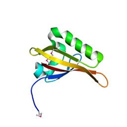
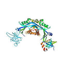
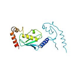
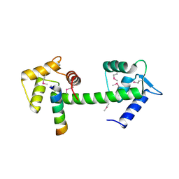
2GGM
DownloadVisualize
BU of 2ggm by Molmil
Human centrin 2 xeroderma pigmentosum group C protein complex
Descriptor: CALCIUM ION, Centrin-2, DNA-repair protein complementing XP-C cells
Authors:Thompson, J.R.
Deposit date:2006-03-24
Release date:2006-04-25
Last modified:2011-07-13
Method:X-RAY DIFFRACTION (2.35 Å)
Cite:The structure of the human centrin 2-xeroderma pigmentosum group C protein complex.
J.Biol.Chem., 281, 2006
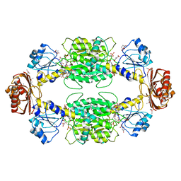
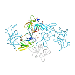
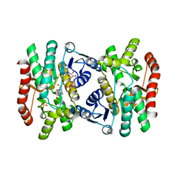
1YBA
DownloadVisualize
BU of 1yba by Molmil
The active form of phosphoglycerate dehydrogenase
Descriptor: 2-OXOGLUTARIC ACID, D-3-phosphoglycerate dehydrogenase, NICOTINAMIDE-ADENINE-DINUCLEOTIDE, ...
Authors:Thompson, J.R, Banaszak, L.J.
Deposit date:2004-12-20
Release date:2005-04-26
Last modified:2024-04-03
Method:X-RAY DIFFRACTION (2.24 Å)
Cite:Vmax Regulation through Domain and Subunit Changes. The Active Form of Phosphoglycerate Dehydrogenase
Biochemistry, 44, 2005
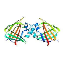
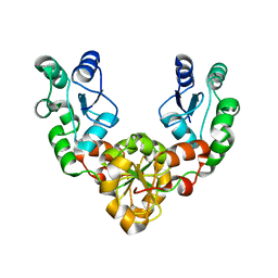
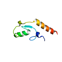
1CBI
DownloadVisualize
BU of 1cbi by Molmil
APO-CELLULAR RETINOIC ACID BINDING PROTEIN I
Descriptor: CELLULAR RETINOIC ACID BINDING PROTEIN I
Authors:Thompson, J.R, Bratt, J.M, Banaszak, L.J.
Deposit date:1995-07-12
Release date:1995-11-14
Last modified:2024-02-07
Method:X-RAY DIFFRACTION (2.7 Å)
Cite:Crystal structure of cellular retinoic acid binding protein I shows increased access to the binding cavity due to formation of an intermolecular beta-sheet.
J.Mol.Biol., 252, 1995
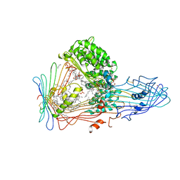
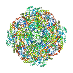
1LSH
DownloadVisualize
BU of 1lsh by Molmil
LIPID-PROTEIN INTERACTIONS IN LIPOVITELLIN
Descriptor: LIPOVITELLIN (LV-1N, LV-1C), LIPOVITELLIN (LV-2), ...
Authors:Thompson, J.R, Banaszak, L.J.
Deposit date:2002-05-17
Release date:2002-07-31
Last modified:2024-04-03
Method:X-RAY DIFFRACTION (1.9 Å)
Cite:Lipid-protein interactions in lipovitellin.
Biochemistry, 41, 2002
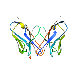
4K07
DownloadVisualize
BU of 4k07 by Molmil
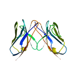
Crystal structure of the amyloid-forming immunoglobulin AL-103 cis-proline 95 mutant
Descriptor: Amyloidogenic immunoglobulin light chain protein AL-103, SULFATE ION
Authors:Thompson, J.R, Berkholz, D.S, Mahlum, E.W, Ramirez-Alvarado, M.
Deposit date:2013-04-03
Release date:2013-10-30
Last modified:2014-01-22
Method:X-RAY DIFFRACTION (2.83 Å)
Cite:Kinetic control in protein folding for light chain amyloidosis and the differential effects of somatic mutations.
J.Mol.Biol., 426, 2014
2Q20
DownloadVisualize
BU of 2q20 by Molmil
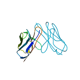
Structure of the germline Vk1 O18/O8 light chain variable domain homodimer
Descriptor: Vk1 O18/O8 germline light chain variable domain
Authors:Thompson, J.R, Ramirez-Alvarado, M, Baden, E.M.
Deposit date:2007-05-25
Release date:2008-04-08
Last modified:2023-08-30
Method:X-RAY DIFFRACTION (1.3 Å)
Cite:Altered dimer interface decreases stability in an amyloidogenic protein.
J.Biol.Chem., 283, 2008
2Q1E
DownloadVisualize
BU of 2q1e by Molmil
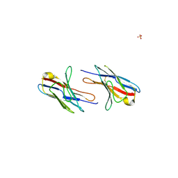
Altered dimer interface decreases stability in an amyloidogenic kappa1 Bence Jones protein.
Descriptor: Amyloidogenic immunoglobulin light chain protein AL-09, SULFATE ION
Authors:Thompson, J.R, Ramirez-Alvarado, M, Baden, E.M.
Deposit date:2007-05-24
Release date:2008-04-08
Last modified:2024-04-03
Method:X-RAY DIFFRACTION (2.55 Å)
Cite:Altered dimer interface decreases stability in an amyloidogenic protein.
J.Biol.Chem., 283, 2008
3DVI
DownloadVisualize
BU of 3dvi by Molmil
Crystal structure of kappa 1 amyloidogenic light chain variable domain
Descriptor: Amyloidogenic light chain variable domain AL-103
Authors:Thompson, J.R, Randles, E.G, Ramirez-Alvarado, M.
Deposit date:2008-07-18
Release date:2009-05-12
Last modified:2023-08-30
Method:X-RAY DIFFRACTION (1.53 Å)
Cite:Structural alterations within native amyloidogenic immunoglobulin light chains.
J.Mol.Biol., 389, 2009
4W8R
DownloadVisualize
BU of 4w8r by Molmil
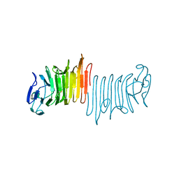
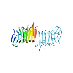
Crystal structure of hemolysin A Y134F from P. mirabilis at 1.5 Angstroms resolution
Descriptor: Hemolysin
Authors:Novak, W.R.P, Glasgow, E, Thompson, J.R, Weaver, T.M.
Deposit date:2014-08-26
Release date:2015-10-07
Last modified:2023-09-27
Method:X-RAY DIFFRACTION (1.519 Å)
Cite:Crystal structure of hemolysin A Y134F from P. mirabilis at 1.5 Angstroms resolution
To Be Published
2B02
DownloadVisualize
BU of 2b02 by Molmil
Crystal Structure of ARNT PAS-B Domain
Descriptor: Aryl hydrocarbon receptor nuclear translocator
Authors:Lee, J, Botuyan, M.V, Nomine, Y, Ohh, M, Thompson, J.R, Mer, G.
Deposit date:2005-09-12
Release date:2006-10-24
Last modified:2021-10-20
Method:X-RAY DIFFRACTION (1.5 Å)
Cite:Crystal Structure and Binding Properties of ARNT PAS-B Heterodimerization Domain
To be Published
2FHD
DownloadVisualize
BU of 2fhd by Molmil
Crystal structure of Crb2 tandem tudor domains
Descriptor: DNA repair protein rhp9/CRB2, PHOSPHATE ION
Authors:Lee, J, Botuyan, M.V, Thompson, J.R, Mer, G.
Deposit date:2005-12-23
Release date:2007-01-02
Last modified:2011-07-13
Method:X-RAY DIFFRACTION (2.4 Å)
Cite:Structural basis for the methylation state-specific recognition of histone H4-K20 by 53BP1 and Crb2 in DNA repair.
Cell(Cambridge,Mass.), 127, 2006
2ETX
DownloadVisualize
BU of 2etx by Molmil
Crystal Structure of MDC1 Tandem BRCT Domains
Descriptor: Mediator of DNA damage checkpoint protein 1
Authors:Wasielewski, E, Kim, Y, Joachimiak, A, Thompson, J.R, Mer, G.
Deposit date:2005-10-27
Release date:2005-11-15
Last modified:2024-02-14
Method:X-RAY DIFFRACTION (1.33 Å)
Cite:Molecular Basis for the Association of Microcephalin (MCPH1) Protein with the Cell Division Cycle Protein 27 (Cdc27) Subunit of the Anaphase-promoting Complex.
J.Biol.Chem., 287, 2012
5KZ5
DownloadVisualize
BU of 5kz5 by Molmil
Architecture of the Human Mitochondrial Iron-Sulfur Cluster Assembly Machinery: the Complex Formed by the Iron Donor, the Sulfur Donor, and the Scaffold
Descriptor: Cysteine desulfurase, mitochondrial, Frataxin, ...
Authors:Gakh, O, Ranatunga, W, Smith, D.Y, Ahlgren, E.C, Al-Karadaghi, S, Thompson, J.R, Isaya, G.
Deposit date:2016-07-22
Release date:2016-08-31
Last modified:2019-12-18
Method:ELECTRON MICROSCOPY (14.3 Å)
Cite:Architecture of the Human Mitochondrial Iron-Sulfur Cluster Assembly Machinery.
J.Biol.Chem., 291, 2016
4W8S
DownloadVisualize
BU of 4w8s by Molmil
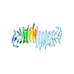
Crystal structure of truncated hemolysin A Q125S/Y134S from P. mirabilis at 1.5 Angstroms resolution
Descriptor: Hemolysin
Authors:Novak, W.R.P, Glasgow, E, Thompson, J.R, Weaver, T.M.
Deposit date:2014-08-26
Release date:2015-10-07
Last modified:2023-09-27
Method:X-RAY DIFFRACTION (1.511 Å)
Cite:Crystal structure of truncated hemolysin A Q125S/Y134S from P. mirabilis at 1.5 Angstroms resolution
To Be Published
4W8T
DownloadVisualize
BU of 4w8t by Molmil
Crystal structure of truncated hemolysin A Q125S from P. mirabilis at 1.5 Angstroms resolution
Descriptor: Hemolysin
Authors:Novak, W.R.P, Glasgow, E, Thompson, J.R, Weaver, T.M.
Deposit date:2014-08-26
Release date:2015-10-07
Last modified:2023-12-27
Method:X-RAY DIFFRACTION (1.539 Å)
Cite:Crystal structure of truncated hemolysin A Q125S from P. mirabilis at 1.5 Angstroms resolution
To Be Published
7RRA
DownloadVisualize
BU of 7rra by Molmil
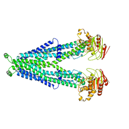
Cryo-EM Structure of Nanodisc reconstituted ABCD1 in inward open conformation
Descriptor: ATP-binding cassette sub-family D member 1
Authors:Alam, A, Le, L.T.M, Thompson, J.R.
Deposit date:2021-08-09
Release date:2022-01-19
Method:ELECTRON MICROSCOPY (4.4 Å)
Cite:Structures of the human peroxisomal fatty acid transporter ABCD1 in a lipid environment
Commun Biol, 5, 2022
7RR9
DownloadVisualize
BU of 7rr9 by Molmil
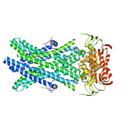
Cryo-EM Structure of Nanodisc reconstituted ABCD1 in nucleotide bound outward open conformation
Descriptor: ATP-binding cassette sub-family D member 1, CHOLESTEROL, MAGNESIUM ION, ...
Authors:Alam, A, Le, L.T.M, Thompson, J.R.
Deposit date:2021-08-09
Release date:2022-01-19
Method:ELECTRON MICROSCOPY (3.5 Å)
Cite:Structures of the human peroxisomal fatty acid transporter ABCD1 in a lipid environment
Commun Biol, 5, 2022
6CO2
DownloadVisualize
BU of 6co2 by Molmil
Structure of an engineered protein (NUDT16TI) in complex with 53BP1 Tudor domains
Descriptor: NUDT16-Tudor-interacting (NUDT16TI), TP53-binding protein 1
Authors:Botuyan, M.V, Thompson, J.R, Cui, G, Mer, G.
Deposit date:2018-03-10
Release date:2018-06-06
Last modified:2023-10-04
Method:X-RAY DIFFRACTION (2.49 Å)
Cite:Mechanism of 53BP1 activity regulation by RNA-binding TIRR and a designer protein.
Nat. Struct. Mol. Biol., 25, 2018
1IB6
DownloadVisualize
BU of 1ib6 by Molmil
CRYSTAL STRUCTURE OF R153C E. COLI MALATE DEHYDROGENASE
Descriptor: MALATE DEHYDROGENASE, NICOTINAMIDE-ADENINE-DINUCLEOTIDE, SULFATE ION
Authors:Bell, J.K, Yennawar, H.P, Wright, S.K, Thompson, J.R, Viola, R.E, Banaszak, L.J.
Deposit date:2001-03-27
Release date:2001-09-19
Last modified:2024-02-07
Method:X-RAY DIFFRACTION (2.1 Å)
Cite:Structural Analyses of a Malate Dehydrogenase with a Variable Active Site
J.Biol.Chem., 276, 2001
3L1X
DownloadVisualize
BU of 3l1x by Molmil
Crystal Structure of U-box Domain of Human E4B Ubiquitin Ligase
Descriptor: Ubiquitin conjugation factor E4 B
Authors:Benirschke, R, Thompson, J.R, Mer, G.
Deposit date:2009-12-14
Release date:2010-05-05
Last modified:2023-09-06
Method:X-RAY DIFFRACTION (2.6 Å)
Cite:Molecular Basis for the Association of Human E4B U Box Ubiquitin Ligase with E2-Conjugating Enzymes UbcH5c and Ubc4.
Structure, 18, 2010
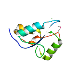
3KTF
DownloadVisualize
BU of 3ktf by Molmil
Structure of the N-terminal BRCT domain of human microcephalin (MCPH1).
Descriptor: CHLORIDE ION, Microcephalin
Authors:Singh, N, Heroux, A, Thompson, J.R, Mer, G.
Deposit date:2009-11-25
Release date:2009-12-15
Last modified:2017-11-01
Method:X-RAY DIFFRACTION (1.6 Å)
Cite:Structure of N-terminal BRCT domain of human microcephalin (MCPH1)
To be Published
3L1Z
DownloadVisualize
BU of 3l1z by Molmil
Crystal structure of the U-BOX domain of human E4B ubiquitin ligase in complex with UBCH5C E2 ubiquitin conjugating enzyme
Descriptor: Ubiquitin conjugation factor E4 B, Ubiquitin-conjugating enzyme E2 D3
Authors:Benirschke, R, Thompson, J.R, Mer, G.
Deposit date:2009-12-14
Release date:2010-05-05
Last modified:2023-09-06
Method:X-RAY DIFFRACTION (3.17 Å)
Cite:Molecular Basis for the Association of Human E4B U Box Ubiquitin Ligase with E2-Conjugating Enzymes UbcH5c and Ubc4.
Structure, 18, 2010
3L1Y
DownloadVisualize
BU of 3l1y by Molmil
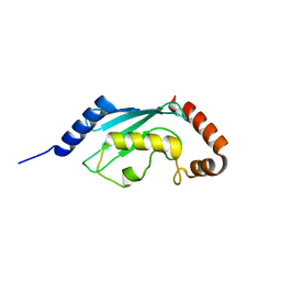
Crystal structure of human UBC4 E2 conjugating enzyme
Descriptor: Ubiquitin-conjugating enzyme E2 D2
Authors:Benirschke, R, Thompson, J.R, Mer, G.
Deposit date:2009-12-14
Release date:2010-05-05
Last modified:2023-09-06
Method:X-RAY DIFFRACTION (1.6 Å)
Cite:Molecular Basis for the Association of Human E4B U Box Ubiquitin Ligase with E2-Conjugating Enzymes UbcH5c and Ubc4.
Structure, 18, 2010
3SZM
DownloadVisualize
BU of 3szm by Molmil
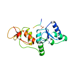
STRUCTURE OF HUMAN MICROCEPHALIN (MCPH1) TANDEM BRCT DOMAINS IN COMPLEX WITH A GAMMA-H2AX PHOSPHOPEPTIDE
Descriptor: Histone H2A.x, Microcephalin
Authors:Singh, N, Thompson, J.R, Mer, G.
Deposit date:2011-07-19
Release date:2011-11-30
Last modified:2012-09-19
Method:X-RAY DIFFRACTION (2.63 Å)
Cite:Dual recognition of phosphoserine and phosphotyrosine in histone variant H2A.X by DNA damage response protein MCPH1.
Proc.Natl.Acad.Sci.USA, 109, 2012
3T1N
DownloadVisualize
BU of 3t1n by Molmil
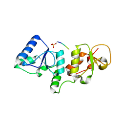
Structure of human MICROCEPHALIN (MCPH1) TANDEM BRCT domains in complex with a CDC27 phosphopeptide
Descriptor: Cdc27 peptide, Microcephalin
Authors:Singh, N, Thompson, J.R, Mer, G.
Deposit date:2011-07-22
Release date:2011-11-30
Last modified:2023-09-13
Method:X-RAY DIFFRACTION (2.6 Å)
Cite:Molecular Basis for the Association of Microcephalin (MCPH1) Protein with the Cell Division Cycle Protein 27 (Cdc27) Subunit of the Anaphase-promoting Complex.
J.Biol.Chem., 287, 2012


1234>

218500

PDB entries from 2024-04-17

PDB statisticsPDBj update infoContact PDBjnumon