Loading
PDBj
MenuPDBj@FacebookPDBj@TwitterPDBj@YouTubewwPDB FoundationwwPDB
RCSB PDBPDBeBMRBAdv. SearchSearch help
Search by PDB author
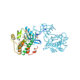
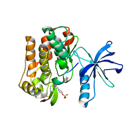
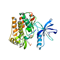
8DWN
DownloadVisualize
BU of 8dwn by Molmil
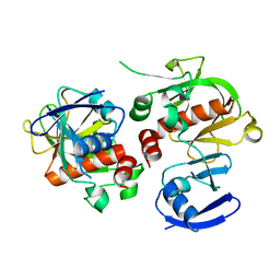
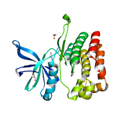
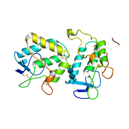
Crystal structure of bis-phosphorylated insulin receptor kinase domain
Descriptor: Insulin receptor subunit beta
Authors:Hubbard, S.R.
Deposit date:2022-08-01
Release date:2023-08-16
Last modified:2023-11-15
Method:X-RAY DIFFRACTION (2.15 Å)
Cite:Crystal structure of bis-phosphorylated insulin receptor kinase domain
To Be Published
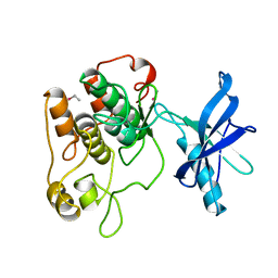
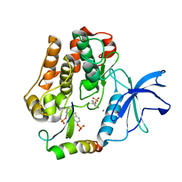
3RGK
DownloadVisualize
BU of 3rgk by Molmil
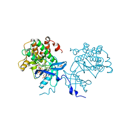
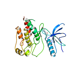
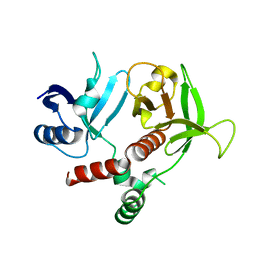
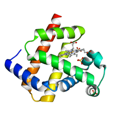
Crystal Structure of Human Myoglobin Mutant K45R
Descriptor: Myoglobin, PROTOPORPHYRIN IX CONTAINING FE, SULFATE ION
Authors:Hubbard, S.R.
Deposit date:2011-04-08
Release date:2011-04-27
Last modified:2023-09-13
Method:X-RAY DIFFRACTION (1.65 Å)
Cite:X-ray crystal structure of a recombinant human myoglobin mutant at 2.8 A resolution
J.Mol.Biol., 20, 1990
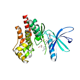
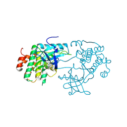
1IR3
DownloadVisualize
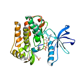
BU of 1ir3 by Molmil
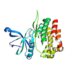
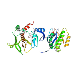
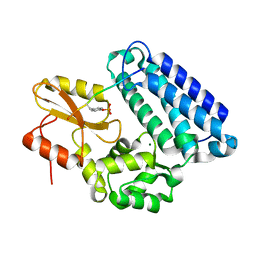
PHOSPHORYLATED INSULIN RECEPTOR TYROSINE KINASE IN COMPLEX WITH PEPTIDE SUBSTRATE AND ATP ANALOG
Descriptor: INSULIN RECEPTOR, MAGNESIUM ION, PEPTIDE SUBSTRATE, ...
Authors:Hubbard, S.R.
Deposit date:1997-09-22
Release date:1998-01-07
Last modified:2023-08-09
Method:X-RAY DIFFRACTION (1.9 Å)
Cite:Crystal structure of the activated insulin receptor tyrosine kinase in complex with peptide substrate and ATP analog.
EMBO J., 16, 1997
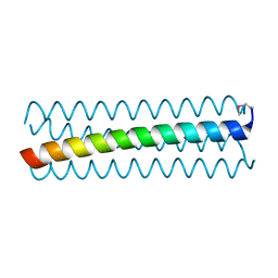
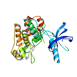
1IRK
DownloadVisualize
BU of 1irk by Molmil
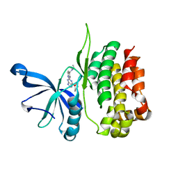
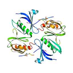
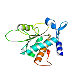
CRYSTAL STRUCTURE OF THE TYROSINE KINASE DOMAIN OF THE HUMAN INSULIN RECEPTOR
Descriptor: ETHYL MERCURY ION, INSULIN RECEPTOR TYROSINE KINASE DOMAIN
Authors:Hubbard, S.R, Wei, L, Ellis, L, Hendrickson, W.A.
Deposit date:1995-01-02
Release date:1995-02-27
Last modified:2024-02-07
Method:X-RAY DIFFRACTION (2.1 Å)
Cite:Crystal structure of the tyrosine kinase domain of the human insulin receptor.
Nature, 372, 1994
7TEU
DownloadVisualize
BU of 7teu by Molmil
Crystal structure of JAK2 JH1 with type II inhibitor YLIU-04-105-1
Descriptor: 3-{(4S)-2-[(cyclopropanecarbonyl)amino]imidazo[1,2-b]pyridazin-6-yl}-N-{3-[(4-ethylpiperazin-1-yl)methyl]-5-(trifluoromethyl)phenyl}-4-methylbenzamide, GLYCEROL, Tyrosine-protein kinase JAK2
Authors:Hubbard, S.R.
Deposit date:2022-01-05
Release date:2023-06-21
Last modified:2023-10-25
Method:X-RAY DIFFRACTION (1.45 Å)
Cite:New scaffolds for type II JAK2 inhibitors overcome the acquired G993A resistance mutation.
Cell Chem Biol, 30, 2023
6D42
DownloadVisualize
BU of 6d42 by Molmil
Crystal structure of the KCa3.1 C-terminal four-helix bundle (with copper)
Descriptor: COPPER (II) ION, Intermediate conductance calcium-activated potassium channel protein 4
Authors:Hubbard, S.R, Ji, T.
Deposit date:2018-04-17
Release date:2018-07-11
Last modified:2019-12-18
Method:X-RAY DIFFRACTION (1.75013649 Å)
Cite:Crystal structure of the C-terminal four-helix bundle of the potassium channel KCa3.1.
PLoS ONE, 13, 2018
3HK0
DownloadVisualize
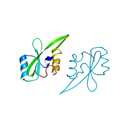
BU of 3hk0 by Molmil
Crystal Structure of the RA and PH Domains of Grb10
Descriptor: Growth factor receptor-bound protein 10, THIOCYANATE ION
Authors:Hubbard, S.R, Depetris, R.S, Wu, J.
Deposit date:2009-05-22
Release date:2009-08-04
Last modified:2024-02-21
Method:X-RAY DIFFRACTION (2.6 Å)
Cite:Structural and functional studies of the Ras-associating and pleckstrin-homology domains of Grb10 and Grb14.
Nat.Struct.Mol.Biol., 16, 2009
4XLV
DownloadVisualize
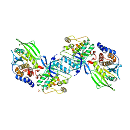
BU of 4xlv by Molmil
Crystal structure of the activated insulin receptor tyrosine kinase dimer
Descriptor: Insulin receptor, MAGNESIUM ION, PHOSPHOMETHYLPHOSPHONIC ACID ADENYLATE ESTER
Authors:Hubbard, S.R, Li, S.
Deposit date:2015-01-13
Release date:2015-03-25
Last modified:2019-12-25
Method:X-RAY DIFFRACTION (2.3 Å)
Cite:The insulin and IGF1 receptor kinase domains are functional dimers in the activated state.
Nat Commun, 6, 2015
8EX1
DownloadVisualize
BU of 8ex1 by Molmil
Crystal structure of JAK2 JH2 (pseudokinase domain) in complex with Reversine
Descriptor: GLYCEROL, N~6~-cyclohexyl-N~2~-(4-morpholin-4-ylphenyl)-9H-purine-2,6-diamine, Tyrosine-protein kinase JAK2
Authors:Sampathkumar, P, Hubbard, S.R.
Deposit date:2022-10-24
Release date:2023-02-01
Last modified:2023-10-25
Method:X-RAY DIFFRACTION (1.5 Å)
Cite:Identification of Novel Small Molecule Ligands for JAK2 Pseudokinase Domain.
Pharmaceuticals, 16, 2023
8EX0
DownloadVisualize
BU of 8ex0 by Molmil
Crystal structure of JAK2 JH2 (pseudokinase domain) in complex with CDK2-IV
Descriptor: 4-{[6-(cyclohexylmethoxy)-7H-purin-2-yl]amino}-N,N-diethylbenzamide, Tyrosine-protein kinase JAK2
Authors:Sampathkumar, P, Hubbard, S.R.
Deposit date:2022-10-24
Release date:2023-02-01
Method:X-RAY DIFFRACTION (1.85 Å)
Cite:Identification of Novel Small Molecule Ligands for JAK2 Pseudokinase Domain.
Pharmaceuticals, 16, 2023
8EX2
DownloadVisualize
BU of 8ex2 by Molmil
Crystal structure of JAK2 JH2 (pseudokinase domain) in complex with HTSA3
Descriptor: 3,5-diphenyl-2-(trifluoromethyl)-6~{H}-pyrazolo[1,5-a]pyrimidin-7-one, GLYCEROL, Tyrosine-protein kinase JAK2
Authors:Sampathkumar, P, Hubbard, S.R.
Deposit date:2022-10-24
Release date:2023-02-01
Last modified:2023-10-25
Method:X-RAY DIFFRACTION (1.9 Å)
Cite:Identification of Novel Small Molecule Ligands for JAK2 Pseudokinase Domain.
Pharmaceuticals, 16, 2023
1AGW
DownloadVisualize
BU of 1agw by Molmil
CRYSTAL STRUCTURE OF THE TYROSINE KINASE DOMAIN OF FIBROBLAST GROWTH FACTOR RECEPTOR 1 IN COMPLEX WITH SU4984 INHIBITOR
Descriptor: 3-[4-(1-FORMYLPIPERAZIN-4-YL)-BENZYLIDENYL]-2-INDOLINONE, FGF RECEPTOR 1
Authors:Mohammadi, M, Schlessinger, J, Hubbard, S.R.
Deposit date:1997-03-25
Release date:1998-03-25
Last modified:2024-02-07
Method:X-RAY DIFFRACTION (2.4 Å)
Cite:Structures of the tyrosine kinase domain of fibroblast growth factor receptor in complex with inhibitors.
Science, 276, 1997
4K81
DownloadVisualize
BU of 4k81 by Molmil
Crystal structure of the Grb14 RA and PH domains in complex with GTP-loaded H-Ras
Descriptor: GLYCEROL, GTPase HRas, GUANOSINE-5'-TRIPHOSPHATE, ...
Authors:Qamra, R, Hubbard, S.R.
Deposit date:2013-04-17
Release date:2013-09-04
Last modified:2023-09-20
Method:X-RAY DIFFRACTION (2.4 Å)
Cite:Structural basis for the interaction of the adaptor protein grb14 with activated ras.
Plos One, 8, 2013
3ML4
DownloadVisualize
BU of 3ml4 by Molmil
Crystal structure of a complex between Dok7 PH-PTB and the MuSK juxtamembrane region
Descriptor: Muscle, skeletal receptor tyrosine-protein kinase, Protein Dok-7
Authors:Bergamin, E, Hubbard, S.R.
Deposit date:2010-04-16
Release date:2010-07-14
Last modified:2011-07-13
Method:X-RAY DIFFRACTION (2.6 Å)
Cite:The Cytoplasmic Adaptor Protein Dok7 Activates the Receptor Tyrosine Kinase MuSK via Dimerization.
Mol.Cell, 39, 2010
3HKL
DownloadVisualize
BU of 3hkl by Molmil
Crystal Structure of the Frizzled-like Cysteine-rich Domain of MuSK
Descriptor: 2-acetamido-2-deoxy-beta-D-glucopyranose-(1-4)-2-acetamido-2-deoxy-beta-D-glucopyranose, Muscle, skeletal receptor tyrosine protein kinase
Authors:Stiegler, A.L, Hubbard, S.R.
Deposit date:2009-05-24
Release date:2009-08-25
Last modified:2020-07-29
Method:X-RAY DIFFRACTION (2.1 Å)
Cite:Crystal structure of the frizzled-like cysteine-rich domain of the receptor tyrosine kinase MuSK.
J.Mol.Biol., 393, 2009
3TCA
DownloadVisualize
BU of 3tca by Molmil
Crystal structure of the Ras-associating and pleckstrin-homology domains of RIAM
Descriptor: Amyloid beta A4 precursor protein-binding family B member 1-interacting protein
Authors:Wu, J, Hubbard, S.R.
Deposit date:2011-08-08
Release date:2012-09-26
Last modified:2023-09-13
Method:X-RAY DIFFRACTION (2.35 Å)
Cite:Rap1-interacting adapter molecule (RIAM) associates with the plasma membrane via a proximity detector.
J.Cell Biol., 199, 2012
1YVH
DownloadVisualize
BU of 1yvh by Molmil
Crystal Structure of the c-Cbl TKB Domain in Complex with the APS pTyr-618 Phosphopeptide
Descriptor: 13-mer fragment of SH2 and PH domain-containing adapter protein APS, CBL E3 ubiquitin protein ligase, MAGNESIUM ION
Authors:Hu, J, Hubbard, S.R.
Deposit date:2005-02-15
Release date:2005-03-08
Last modified:2023-11-15
Method:X-RAY DIFFRACTION (2.05 Å)
Cite:Structural Characterization of a Novel Cbl Phosphotyrosine Recognition Motif in the APS Family of Adapter Proteins
J.Biol.Chem., 280, 2005
3BCH
DownloadVisualize
BU of 3bch by Molmil
Crystal Structure of the Human Laminin Receptor Precursor
Descriptor: 40S ribosomal protein SA
Authors:Jamieson, K.V, Wu, J, Hubbard, S.R, Meruelo, D.
Deposit date:2007-11-12
Release date:2007-12-04
Last modified:2024-02-21
Method:X-RAY DIFFRACTION (2.15 Å)
Cite:Crystal structure of the human laminin receptor precursor.
J.Biol.Chem., 283, 2008
3BU3
DownloadVisualize
BU of 3bu3 by Molmil
Crystal structure of the insulin receptor kinase in complex with IRS2 KRLB peptide
Descriptor: Insulin receptor substrate 2, insulin receptor subunit beta
Authors:Wu, J, Hubbard, S.R.
Deposit date:2007-12-31
Release date:2008-02-19
Last modified:2021-10-20
Method:X-RAY DIFFRACTION (1.65 Å)
Cite:Structural and biochemical characterization of the KRLB region in insulin receptor substrate-2.
Nat.Struct.Mol.Biol., 15, 2008
3BU5
DownloadVisualize
BU of 3bu5 by Molmil
Crystal structure of the insulin receptor kinase in complex with IRS2 KRLB peptide and ATP
Descriptor: ADENOSINE-5'-TRIPHOSPHATE, Insulin receptor substrate 2, MAGNESIUM ION, ...
Authors:Wu, J, Hubbard, S.R.
Deposit date:2007-12-31
Release date:2008-02-19
Last modified:2021-10-20
Method:X-RAY DIFFRACTION (2.1 Å)
Cite:Structural and biochemical characterization of the KRLB region in insulin receptor substrate-2.
Nat.Struct.Mol.Biol., 15, 2008
2B4S
DownloadVisualize
BU of 2b4s by Molmil
Crystal structure of a complex between PTP1B and the insulin receptor tyrosine kinase
Descriptor: Insulin receptor, SULFATE ION, Tyrosine-protein phosphatase, ...
Authors:Li, S, Depetris, R.S, Barford, D, Chernoff, J, Hubbard, S.R.
Deposit date:2005-09-26
Release date:2005-11-15
Last modified:2023-11-15
Method:X-RAY DIFFRACTION (2.3 Å)
Cite:Crystal Structure of a Complex between Protein Tyrosine Phosphatase 1B and the Insulin Receptor Tyrosine Kinase.
Structure, 13, 2005
2AUG
DownloadVisualize
BU of 2aug by Molmil
Crystal structure of the Grb14 SH2 domain
Descriptor: Growth factor receptor-bound protein 14
Authors:Depetris, R.S, Hu, J, Gimpelevich, I, Holt, L.J, Daly, R.J, Hubbard, S.R.
Deposit date:2005-08-27
Release date:2005-11-01
Last modified:2023-08-23
Method:X-RAY DIFFRACTION (2.3 Å)
Cite:Structural basis for inhibition of the insulin receptor by the adaptor protein grb14.
Mol.Cell, 20, 2005
3BU6
DownloadVisualize
BU of 3bu6 by Molmil
Crystal structure of the insulin receptor kinase in complex with IRS2 KRLB phosphopeptide
Descriptor: Insulin receptor substrate 2, insulin receptor subunit beta
Authors:Wu, J, Hubbard, S.R.
Deposit date:2007-12-31
Release date:2008-02-19
Last modified:2021-10-20
Method:X-RAY DIFFRACTION (1.95 Å)
Cite:Structural and biochemical characterization of the KRLB region in insulin receptor substrate-2.
Nat.Struct.Mol.Biol., 15, 2008
2AUH
DownloadVisualize
BU of 2auh by Molmil
Crystal structure of the Grb14 BPS region in complex with the insulin receptor tyrosine kinase
Descriptor: CALCIUM ION, Growth factor receptor-bound protein 14, Insulin receptor
Authors:Depetris, R.S, Hu, J, Gimpelevich, I, Holt, L.J, Daly, R.J, Hubbard, S.R.
Deposit date:2005-08-27
Release date:2005-11-01
Last modified:2023-11-15
Method:X-RAY DIFFRACTION (3.2 Å)
Cite:Structural basis for inhibition of the insulin receptor by the adaptor protein grb14.
Mol.Cell, 20, 2005
3D94
DownloadVisualize
BU of 3d94 by Molmil
Crystal structure of the insulin-like growth factor-1 receptor kinase in complex with PQIP
Descriptor: 3-[cis-3-(4-methylpiperazin-1-yl)cyclobutyl]-1-(2-phenylquinolin-7-yl)imidazo[1,5-a]pyrazin-8-amine, CALCIUM ION, Insulin-like growth factor 1 receptor beta chain
Authors:Wu, J, Li, W, Miller, W.T, Hubbard, S.R.
Deposit date:2008-05-26
Release date:2008-07-29
Last modified:2023-08-30
Method:X-RAY DIFFRACTION (2.3 Å)
Cite:Small-molecule inhibition and activation-loop trans-phosphorylation of the IGF1 receptor
Embo J., 27, 2008


123>

218853

PDB entries from 2024-04-24

PDB statisticsPDBj update infoContact PDBjnumon