Loading
PDBj
MenuPDBj@FacebookPDBj@TwitterPDBj@YouTubewwPDB FoundationwwPDB
RCSB PDBPDBeBMRBAdv. SearchSearch help
Search by PDB author
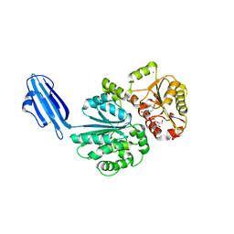
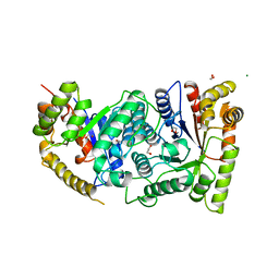
1BRL
DownloadVisualize
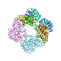
BU of 1brl by Molmil
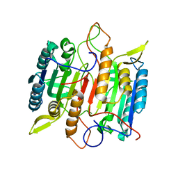
THREE-DIMENSIONAL STRUCTURE OF BACTERIAL LUCIFERASE FROM VIBRIO HARVEYI AT 2.4 ANGSTROMS RESOLUTION
Descriptor: BACTERIAL LUCIFERASE, PHOSPHATE ION
Authors:Fisher, A.J, Rayment, I.
Deposit date:1995-03-20
Release date:1996-04-01
Last modified:2024-02-07
Method:X-RAY DIFFRACTION (2.4 Å)
Cite:Three-dimensional structure of bacterial luciferase from Vibrio harveyi at 2.4 A resolution.
Biochemistry, 34, 1995
1LUC
DownloadVisualize
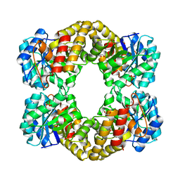
BU of 1luc by Molmil
BACTERIAL LUCIFERASE
Descriptor: 1,2-ETHANEDIOL, BACTERIAL LUCIFERASE, MAGNESIUM ION
Authors:Fisher, A.J, Rayment, I.
Deposit date:1996-05-10
Release date:1996-12-07
Last modified:2024-02-14
Method:X-RAY DIFFRACTION (1.5 Å)
Cite:The 1.5-A resolution crystal structure of bacterial luciferase in low salt conditions.
J.Biol.Chem., 271, 1996
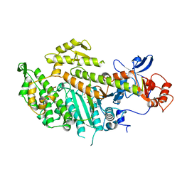
1MMD
DownloadVisualize
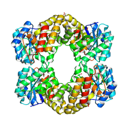
BU of 1mmd by Molmil
TRUNCATED HEAD OF MYOSIN FROM DICTYOSTELIUM DISCOIDEUM COMPLEXED WITH MGADP-BEF3
Descriptor: ADENOSINE-5'-DIPHOSPHATE, BERYLLIUM TRIFLUORIDE ION, MAGNESIUM ION, ...
Authors:Fisher, A.J, Holden, H.M, Rayment, I.
Deposit date:1995-03-21
Release date:1996-08-17
Last modified:2024-02-14
Method:X-RAY DIFFRACTION (2 Å)
Cite:X-ray structures of the myosin motor domain of Dictyostelium discoideum complexed with MgADP.BeFx and MgADP.AlF4-.
Biochemistry, 34, 1995
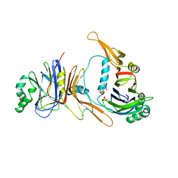
1P35
DownloadVisualize
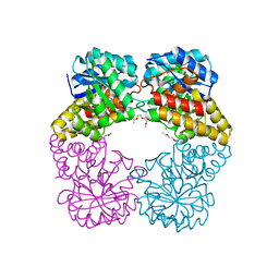
BU of 1p35 by Molmil
CRYSTAL STRUCTURE OF BACULOVIRUS P35
Descriptor: 1,2-ETHANEDIOL, P35, PHOSPHATE ION
Authors:Fisher, A.J, Delacruz, W.P, Zoog, S.J, Schneider, C.L, Friesen, P.D.
Deposit date:1998-09-18
Release date:1999-06-01
Last modified:2024-02-14
Method:X-RAY DIFFRACTION (2.2 Å)
Cite:Crystal structure of baculovirus P35: role of a novel reactive site loop in apoptotic caspase inhibition.
EMBO J., 18, 1999
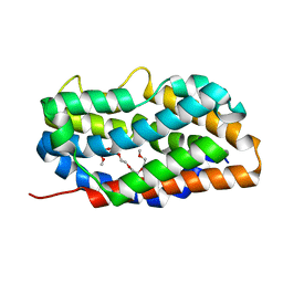
4RAJ
DownloadVisualize
BU of 4raj by Molmil
Crystal structure of heme oxygenase 2 from Chlamydomonas reinhardtii without heme.
Descriptor: 1-(2-METHOXY-ETHOXY)-2-{2-[2-(2-METHOXY-ETHOXY]-ETHOXY}-ETHANE, Heme oxygenase
Authors:Fisher, A.J, Lagarias, J.C.
Deposit date:2014-09-10
Release date:2015-09-16
Last modified:2023-09-20
Method:X-RAY DIFFRACTION (1.84 Å)
Cite:Crystal structure of heme oxygenase 2 from Chlamydomonas reinhardtii without heme.
To be Published
4R84
DownloadVisualize
BU of 4r84 by Molmil
Crystal structure of Sialyltransferase from Photobacterium damsela with CMP-3F(a)Neu5Ac bound
Descriptor: CALCIUM ION, CYTIDINE-5'-MONOPHOSPHATE-3-FLUORO-N-ACETYL-NEURAMINIC ACID, Sialyltransferase 0160
Authors:Fisher, A.J, Chen, X, Li, Y, Huynh, N.
Deposit date:2014-08-29
Release date:2014-12-03
Last modified:2023-09-20
Method:X-RAY DIFFRACTION (1.7 Å)
Cite:Crystal structures of sialyltransferase from Photobacterium damselae.
Febs Lett., 588, 2014
7TNI
DownloadVisualize
BU of 7tni by Molmil
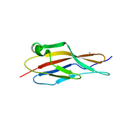
Structure of EC12 Y1392W variant of BT-R1 from Manduca sexta, a Cry1A toxin binding domain
Descriptor: Cadherin, MAGNESIUM ION
Authors:Fisher, A.J, Wilcox, X.E.
Deposit date:2022-01-21
Release date:2022-05-04
Last modified:2023-10-18
Method:X-RAY DIFFRACTION (1.9 Å)
Cite:Functional and Structural Analysis of the Toxin-Binding Site of the Cadherin G-Protein-Coupled Receptor, BT-R 1 , for Cry1A Toxins of Bacillus thuringiensis .
Biochemistry, 61, 2022
8E4X
DownloadVisualize
BU of 8e4x by Molmil
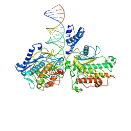
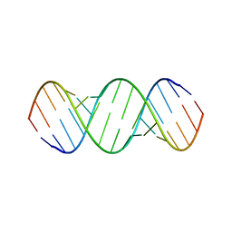
Human Adenosine Deaminase Acting on dsRNA (ADAR2-R2D) bound to dsRNA containing a G:3-deaza dA pair adjacent to the target site
Descriptor: Double-stranded RNA-specific editase 1, INOSITOL HEXAKISPHOSPHATE, RNA (5-R(*GP*CP*UP*CP*GP*CP*GP*AP*UP*GP*CP*GP*(8AZ)P*GP*AP*GP*GP*GP*CP* UP*CP*UP*GP*AP*UP*AP*GP*CP*UP*AP*CP*G)-3), ...
Authors:Fisher, A.J, Mendoza, H.G.
Deposit date:2022-08-19
Release date:2022-10-26
Last modified:2023-10-18
Method:X-RAY DIFFRACTION (2.8 Å)
Cite:ADAR activation by inducing a syn conformation at guanosine adjacent to an editing site.
Nucleic Acids Res., 50, 2022
6VLB
DownloadVisualize
BU of 6vlb by Molmil
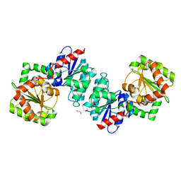
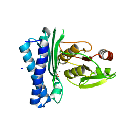
Crystal structure of ligand-free UDP-GlcNAc 2-epimerase from Neisseria meningitidis
Descriptor: 1,2-ETHANEDIOL, SODIUM ION, UDP-N-acetylglucosamine 2-epimerase
Authors:Fisher, A.J, Hurlburt, N.K.
Deposit date:2020-01-23
Release date:2020-11-11
Last modified:2023-10-11
Method:X-RAY DIFFRACTION (1.85 Å)
Cite:Structural characterization of a nonhydrolyzing UDP-GlcNAc 2-epimerase from Neisseria meningitidis serogroup A.
Acta Crystallogr.,Sect.F, 76, 2020
6VLC
DownloadVisualize
BU of 6vlc by Molmil
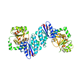
Crystal structure of UDP-GlcNAc 2-epimerase from Neisseria meningitidis bound to UDP-GlcNAc
Descriptor: UDP-N-acetylglucosamine 2-epimerase, URIDINE-DIPHOSPHATE-N-ACETYLGLUCOSAMINE
Authors:Fisher, A.J, Hurlburt, N.K.
Deposit date:2020-01-23
Release date:2020-11-11
Last modified:2023-10-11
Method:X-RAY DIFFRACTION (2.15 Å)
Cite:Structural characterization of a nonhydrolyzing UDP-GlcNAc 2-epimerase from Neisseria meningitidis serogroup A.
Acta Crystallogr.,Sect.F, 76, 2020
5DJI
DownloadVisualize
BU of 5dji by Molmil
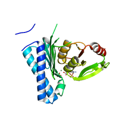
Structure of M. tuberculosis CysQ, a PAP phosphatase with AMP, PO4, and 2Mg bound
Descriptor: 3'-phosphoadenosine 5'-phosphate phosphatase, ADENOSINE MONOPHOSPHATE, MAGNESIUM ION, ...
Authors:Fisher, A.J, Erickson, A.I.
Deposit date:2015-09-02
Release date:2015-11-11
Last modified:2023-09-27
Method:X-RAY DIFFRACTION (1.659 Å)
Cite:Crystal Structures of Mycobacterium tuberculosis CysQ, with Substrate and Products Bound.
Biochemistry, 54, 2015
5DJG
DownloadVisualize
BU of 5djg by Molmil
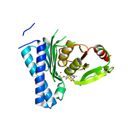
Structure of M. tuberculosis CysQ, a PAP phosphatase with PAP, Mg, and Li bound
Descriptor: 3'-phosphoadenosine 5'-phosphate phosphatase, ADENOSINE-3'-5'-DIPHOSPHATE, LITHIUM ION, ...
Authors:Fisher, A.J, Erickson, A.I.
Deposit date:2015-09-02
Release date:2015-11-11
Last modified:2023-09-27
Method:X-RAY DIFFRACTION (1.951 Å)
Cite:Crystal Structures of Mycobacterium tuberculosis CysQ, with Substrate and Products Bound.
Biochemistry, 54, 2015
5DJH
DownloadVisualize
BU of 5djh by Molmil
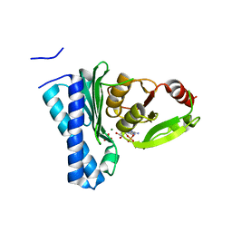
Structure of M. tuberculosis CysQ, a PAP phosphatase with AMP, PO4, and 3Mg bound
Descriptor: 3'-phosphoadenosine 5'-phosphate phosphatase, ADENOSINE MONOPHOSPHATE, MAGNESIUM ION, ...
Authors:Fisher, A.J, Erickson, A.I.
Deposit date:2015-09-02
Release date:2015-11-11
Last modified:2023-09-27
Method:X-RAY DIFFRACTION (1.451 Å)
Cite:Crystal Structures of Mycobacterium tuberculosis CysQ, with Substrate and Products Bound.
Biochemistry, 54, 2015
5DJJ
DownloadVisualize
BU of 5djj by Molmil
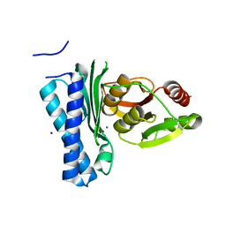
Structure of M. tuberculosis CysQ, a PAP phosphatase with PO4 and 2Mg bound
Descriptor: 3'-phosphoadenosine 5'-phosphate phosphatase, MAGNESIUM ION, PHOSPHATE ION, ...
Authors:Fisher, A.J, Erickson, A.I.
Deposit date:2015-09-02
Release date:2015-11-11
Last modified:2023-09-27
Method:X-RAY DIFFRACTION (1.501 Å)
Cite:Crystal Structures of Mycobacterium tuberculosis CysQ, with Substrate and Products Bound.
Biochemistry, 54, 2015
5DJK
DownloadVisualize
BU of 5djk by Molmil
Structure of M. tuberculosis CysQ, a PAP phosphatase with PO4 and 2Ca bound
Descriptor: 3'-phosphoadenosine 5'-phosphate phosphatase, CALCIUM ION, PHOSPHATE ION, ...
Authors:Fisher, A.J, Erickson, A.I.
Deposit date:2015-09-02
Release date:2015-11-11
Last modified:2023-09-27
Method:X-RAY DIFFRACTION (1.799 Å)
Cite:Crystal Structures of Mycobacterium tuberculosis CysQ, with Substrate and Products Bound.
Biochemistry, 54, 2015
6BGB
DownloadVisualize
BU of 6bgb by Molmil
Crystal Structure of the 16mer GCAGNCUUAAGUCUGC containing BrPh 7-triazolyl-8-aza-7-deazaadenosine
Descriptor: (5'-R(*GP*CP*AP*GP*(A7C)P*CP*UP*UP*AP*AP*GP*UP*CP*UP*GP*C)-3')
Authors:Fisher, A.J, Beal, P.A, Mumbleau, M.M.
Deposit date:2017-10-27
Release date:2017-12-06
Last modified:2024-03-13
Method:X-RAY DIFFRACTION (1.65 Å)
Cite:Controlling miRNA-like off-target effects of an siRNA with nucleobase modifications.
Org. Biomol. Chem., 15, 2017
6DWO
DownloadVisualize
BU of 6dwo by Molmil
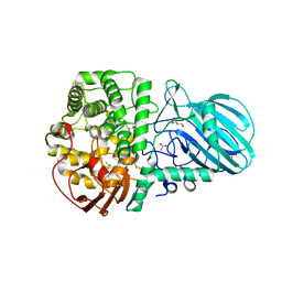
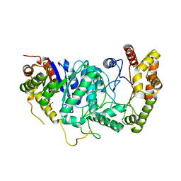
Crystal structure of alpha-1-2-mannosidase from Enterococcus faecalis V583
Descriptor: ACETATE ION, Alpha-1,2-mannosidase, CALCIUM ION, ...
Authors:Fisher, A.J, Li, Y.
Deposit date:2018-06-26
Release date:2019-10-30
Last modified:2023-10-11
Method:X-RAY DIFFRACTION (2.15 Å)
Cite:Enterococcus faecalis alpha 1-2-mannosidase (EfMan-I): an efficient catalyst for glycoprotein N-glycan modification.
Febs Lett., 594, 2020
4IME
DownloadVisualize
BU of 4ime by Molmil
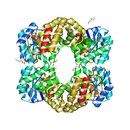
Crystal Structure of Pasteurella multocida N-Acetyl-D-Neuraminic acid lyase K164A Mutant
Descriptor: 1-(2-METHOXY-ETHOXY)-2-{2-[2-(2-METHOXY-ETHOXY]-ETHOXY}-ETHANE, 1-ETHOXY-2-(2-METHOXYETHOXY)ETHANE, N-acetylneuraminate lyase
Authors:Fisher, A.J, Huynh, N.
Deposit date:2013-01-02
Release date:2013-11-06
Last modified:2024-02-28
Method:X-RAY DIFFRACTION (1.75 Å)
Cite:Structural Basis for Substrate Specificity and Mechanism of N-Acetyl-d-neuraminic Acid Lyase from Pasteurella multocida.
Biochemistry, 52, 2013
4IMG
DownloadVisualize
BU of 4img by Molmil
Crystal Structure of Pasteurella multocida N-Acetyl-D-Neuraminic acid lyase K164 mutant complexed with N-Glycolylneuraminic acid
Descriptor: 1-ETHOXY-2-(2-METHOXYETHOXY)ETHANE, 3,5-dideoxy-5-[(hydroxyacetyl)amino]-D-glycero-D-galacto-non-2-ulosonic acid, N-acetylneuraminate lyase
Authors:Fisher, A.J, Huynh, N.
Deposit date:2013-01-02
Release date:2013-11-06
Last modified:2024-02-28
Method:X-RAY DIFFRACTION (1.85 Å)
Cite:Structural Basis for Substrate Specificity and Mechanism of N-Acetyl-d-neuraminic Acid Lyase from Pasteurella multocida.
Biochemistry, 52, 2013
4IMC
DownloadVisualize
BU of 4imc by Molmil
Crystal Structure of Pasteurella multocida N-Acetyl-D-Neuraminic acid lyase
Descriptor: 1,2-ETHANEDIOL, N-acetylneuraminate lyase, PHOSPHATE ION
Authors:Fisher, A.J, Huynh, N.
Deposit date:2013-01-02
Release date:2013-11-06
Last modified:2024-02-28
Method:X-RAY DIFFRACTION (1.85 Å)
Cite:Structural Basis for Substrate Specificity and Mechanism of N-Acetyl-d-neuraminic Acid Lyase from Pasteurella multocida.
Biochemistry, 52, 2013
4IMD
DownloadVisualize
BU of 4imd by Molmil
Crystal Structure of Pasteurella multocida N-Acetyl-D-Neuraminic acid lyase trapped with pyruvate covalently bound through a Schiff base to Lys164
Descriptor: 1,2-ETHANEDIOL, N-acetylneuraminate lyase, PHOSPHATE ION
Authors:Fisher, A.J, Huynh, N.
Deposit date:2013-01-02
Release date:2013-11-06
Last modified:2013-12-11
Method:X-RAY DIFFRACTION (2.1 Å)
Cite:Structural Basis for Substrate Specificity and Mechanism of N-Acetyl-d-neuraminic Acid Lyase from Pasteurella multocida.
Biochemistry, 52, 2013
4IMF
DownloadVisualize
BU of 4imf by Molmil
Crystal Structure of Pasteurella multocida N-Acetyl-D-Neuraminic acid lyase K164 mutant complexed with N-Acetylneuraminic acid
Descriptor: 1-ETHOXY-2-(2-METHOXYETHOXY)ETHANE, 5-(acetylamino)-3,5-dideoxy-D-glycero-D-galacto-non-2-ulosonic acid, CHLORIDE ION, ...
Authors:Fisher, A.J, Huynh, N.
Deposit date:2013-01-02
Release date:2013-11-06
Last modified:2024-02-28
Method:X-RAY DIFFRACTION (1.9 Å)
Cite:Structural Basis for Substrate Specificity and Mechanism of N-Acetyl-d-neuraminic Acid Lyase from Pasteurella multocida.
Biochemistry, 52, 2013
2NN3
DownloadVisualize
BU of 2nn3 by Molmil
structure of pro-sf-caspase-1
Descriptor: Caspase-1
Authors:Fisher, A.J, Ni, L.
Deposit date:2006-10-23
Release date:2007-10-23
Last modified:2023-08-30
Method:X-RAY DIFFRACTION (3 Å)
Cite:Pro-sfcapsase-1, structural insights into activation mechanism of caspases
To be Published
7KL5
DownloadVisualize
BU of 7kl5 by Molmil
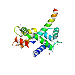
Structure of Calmodulin Bound to the Cardiac Ryanodine Receptor (RyR2) at Residues: Phe4246 to Val4271
Descriptor: 1,2-ETHANEDIOL, CALCIUM ION, Calmodulin-1, ...
Authors:Fisher, A.J, Ames, J.B, Yu, Q.
Deposit date:2020-10-29
Release date:2021-04-07
Last modified:2023-10-18
Method:X-RAY DIFFRACTION (1.65 Å)
Cite:The Crystal Structure of Calmodulin Bound to the Cardiac Ryanodine Receptor (RyR2) at Residues Phe4246-Val4271 Reveals a Fifth Calcium Binding Site.
Biochemistry, 60, 2021
4R37
DownloadVisualize
BU of 4r37 by Molmil
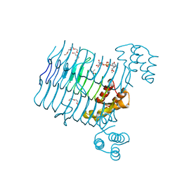
Crystal structure analysis of LpxA, a UDP-N-acetylglucosamine acyltransferase from Bacteroides fragilis 9343 with UDP-GlcNAc
Descriptor: 2-(2-METHOXYETHOXY)ETHANOL, 3,6,9,12,15,18,21,24-OCTAOXAHEXACOSAN-1-OL, ACETATE ION, ...
Authors:Fisher, A.J, Chen, X, Ngo, A.
Deposit date:2014-08-14
Release date:2015-05-06
Last modified:2023-09-20
Method:X-RAY DIFFRACTION (1.9 Å)
Cite:Structures of Bacteroides fragilis uridine 5'-diphosphate-N-acetylglucosamine (UDP-GlcNAc) acyltransferase (BfLpxA).
Acta Crystallogr.,Sect.D, 71, 2015

218500

PDB entries from 2024-04-17

PDB statisticsPDBj update infoContact PDBjnumon