Loading
PDBj
MenuPDBj@FacebookPDBj@TwitterPDBj@YouTubewwPDB FoundationwwPDB
RCSB PDBPDBeBMRBAdv. SearchSearch help
Search by PDB author
1CFG
DownloadVisualize
BU of 1cfg by Molmil
MEMBRANE-BINDING PEPTIDE FROM THE C2 DOMAIN OF FACTOR VIII FORMS AN AMPHIPATHIC STRUCTURE AS DETERMINED BY NMR SPECTROSCOPY
Descriptor: COAGULATION FACTOR VIII
Authors:Baleja, J.D, Gilbert, G.E.
Deposit date:1994-11-10
Release date:1995-11-02
Last modified:2022-02-16
Method:SOLUTION NMR
Cite:Membrane-binding peptide from the C2 domain of factor VIII forms an amphipathic structure as determined by NMR spectroscopy.
Biochemistry, 34, 1995
3BN6
DownloadVisualize
BU of 3bn6 by Molmil
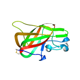
Crystal Structure of the C2 Domain of Bovine Lactadherin at 1.67 Angstrom Resolution
Descriptor: Lactadherin
Authors:Shao, C, Novakovic, V.A, Head, J.F, Seaton, B.A, Gilbert, G.E.
Deposit date:2007-12-13
Release date:2007-12-25
Last modified:2023-09-20
Method:X-RAY DIFFRACTION (1.67 Å)
Cite:Crystal structure of lactadherin C2 domain at 1.7A resolution with mutational and computational analyses of its membrane-binding motif.
J.Biol.Chem., 283, 2008
1FAC
DownloadVisualize
BU of 1fac by Molmil
COAGULATION FACTOR VIII, NMR, 1 STRUCTURE
Descriptor: COAGULATION FACTOR VIII
Authors:Veeraraghavan, S, Baleja, J.D, Gilbert, G.E.
Deposit date:1996-06-30
Release date:1997-01-11
Last modified:2022-02-23
Method:SOLUTION NMR
Cite:Structure and topography of the membrane-binding C2 domain of factor VIII in the presence of dodecylphosphocholine micelles.
Biochem.J., 332, 1998

218196

PDB entries from 2024-04-10

PDB statisticsPDBj update infoContact PDBjnumon