2J1D
 
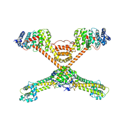
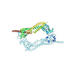 | | Crystallization of hDaam1 C-terminal Fragment | | 分子名称: | DISHEVELED-ASSOCIATED ACTIVATOR OF MORPHOGENESIS 1, GLYCEROL, PHOSPHATE ION | | 著者 | Lu, J, Meng, W, Poy, F, Eck, M.J. | | 登録日 | 2006-08-10 | | 公開日 | 2007-05-15 | | 最終更新日 | 2011-07-13 | | 実験手法 | X-RAY DIFFRACTION (2.25 Å) | | 主引用文献 | Structure of the Fh2 Domain of Daam1: Implications for Formin Regulation of Actin Assembly.
J.Mol.Biol., 369, 2007
|
|
2Z6E
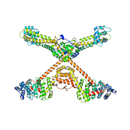
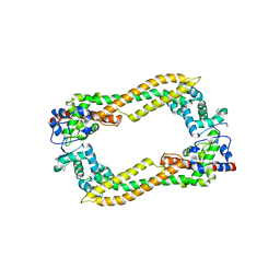 | | Crystal Structure of Human DAAM1 FH2 | | 分子名称: | Disheveled-associated activator of morphogenesis 1 | | 著者 | Yamashita, M, Higashi, T, Sato, Y, Shirakawa, R, Kita, T, Horiuchi, H, Fukai, S, Nureki, O. | | 登録日 | 2007-07-31 | | 公開日 | 2008-05-27 | | 最終更新日 | 2024-03-13 | | 実験手法 | X-RAY DIFFRACTION (2.8 Å) | | 主引用文献 | Crystal structure of human DAAM1 formin homology 2 domain
Genes Cells, 12, 2007
|
|
1UX4
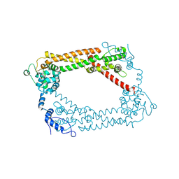
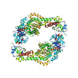
 | | Crystal structures of a Formin Homology-2 domain reveal a tethered-dimer architecture | | 分子名称: | BNI1 PROTEIN | | 著者 | Xu, Y, Moseley, J.B, Sagot, I, Poy, F, Pellman, D, Goode, B.L, Eck, M.J. | | 登録日 | 2004-02-19 | | 公開日 | 2004-03-11 | | 最終更新日 | 2019-05-08 | | 実験手法 | X-RAY DIFFRACTION (3.3 Å) | | 主引用文献 | Crystal Structures of a Formin Homology-2 Domain Reveal a Tethered Dimer Architecture
Cell(Cambridge,Mass.), 116, 2004
|
|
1V9D
 | | Crystal structure of the core FH2 domain of mouse mDia1 | | 分子名称: | Diaphanous protein homolog 1, SULFATE ION | | 著者 | Shimada, A, Nyitrai, M, Vetter, I.R, Kuhlmann, D, Bugyi, B, Narumiya, S, Geeves, M.A, Wittinghofer, A. | | 登録日 | 2004-01-24 | | 公開日 | 2004-03-09 | | 最終更新日 | 2023-12-27 | | 実験手法 | X-RAY DIFFRACTION (2.6 Å) | | 主引用文献 | The core FH2 domain of diaphanous-related formins is an elongated actin binding protein that inhibits polymerization.
Mol.Cell, 13, 2004
|
|
1UX5
 | | Crystal Structures of a Formin Homology-2 domain reveal a flexibly tethered dimer architecture | | 分子名称: | BNI1 PROTEIN | | 著者 | Xu, Y, Moseley, J.B, Sagot, I, Poy, F, Pellman, D, Goode, B.L, Eck, M.J. | | 登録日 | 2004-02-19 | | 公開日 | 2004-03-11 | | 最終更新日 | 2019-05-08 | | 実験手法 | X-RAY DIFFRACTION (2.5 Å) | | 主引用文献 | Crystal Structures of a Formin Homology-2 Domain Reveal a Tethered Dimer Architecture
Cell(Cambridge,Mass.), 116, 2004
|
|
3O4X
 | |
3OBV
 | |
4EAH
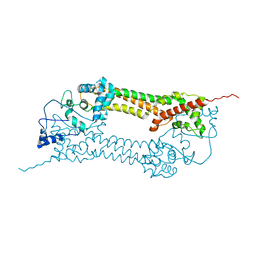
 | | Crystal structure of the formin homology 2 domain of FMNL3 bound to actin | | 分子名称: | ACETATE ION, ADENOSINE-5'-TRIPHOSPHATE, Actin, ... | | 著者 | Thompson, M.E, Heimsath, E.G, Gauvin, T.J, Higgs, H.N, Kull, F.J. | | 登録日 | 2012-03-22 | | 公開日 | 2012-12-12 | | 最終更新日 | 2024-02-28 | | 実験手法 | X-RAY DIFFRACTION (3.4 Å) | | 主引用文献 | FMNL3 FH2-actin structure gives insight into formin-mediated actin nucleation and elongation.
Nat.Struct.Mol.Biol., 20, 2013
|
|
1Y64
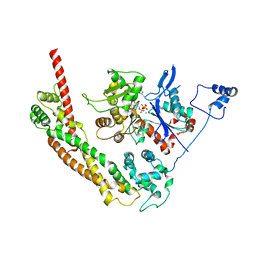 | | Bni1p Formin Homology 2 Domain complexed with ATP-actin | | 分子名称: | ADENOSINE-5'-TRIPHOSPHATE, Actin, alpha skeletal muscle, ... | | 著者 | Otomo, T, Tomchick, D.R, Otomo, C, Panchal, S.C, Machius, M, Rosen, M.K. | | 登録日 | 2004-12-03 | | 公開日 | 2005-01-18 | | 最終更新日 | 2011-07-13 | | 実験手法 | X-RAY DIFFRACTION (3.05 Å) | | 主引用文献 | Structural basis of actin filament nucleation and processive capping by a formin homology 2 domain
Nature, 433, 2005
|
|


