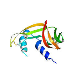
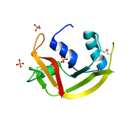
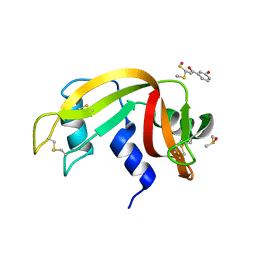
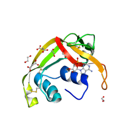
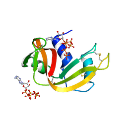
4X09
 
 | | Structure of human RNase 6 in complex with sulphate anions | | 分子名称: | GLYCEROL, Ribonuclease K6, SULFATE ION | | 著者 | Prats-Ejarque, G, Arranz-Trullen, J, Blanco, J.A, Pulido, D, Moussaoui, M, Boix, E. | | 登録日 | 2014-11-21 | | 公開日 | 2016-04-06 | | 最終更新日 | 2024-01-10 | | 実験手法 | X-RAY DIFFRACTION (1.722 Å) | | 主引用文献 | The first crystal structure of human RNase 6 reveals a novel substrate-binding and cleavage site arrangement.
Biochem.J., 473, 2016
|
|
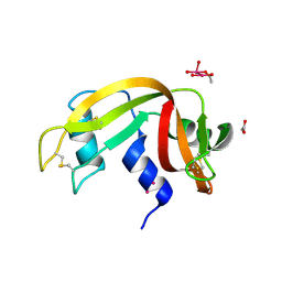
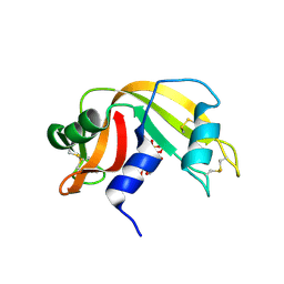
8C3B
 | | X-ray structure of RNase A upon reaction with a Ruthenium(II)-arene Complexed with Glycosylated Carbene Ligands (5) | | 分子名称: | (1,3-dimethyl-2~{H}-imidazol-2-yl)-oxidanyl-oxidanylidene-ruthenium, (1,3-dimethylimidazol-1-ium-2-yl)-tetrakis(oxidanyl)ruthenium, (1,3-dimethylimidazol-1-ium-2-yl)-tris(oxidanyl)ruthenium, ... | | 著者 | Ferraro, G, Merlino, A. | | 登録日 | 2022-12-23 | | 公開日 | 2024-01-10 | | 実験手法 | X-RAY DIFFRACTION (1.24 Å) | | 主引用文献 | Ruthenium(II)-arene Complexes with Glycosylated Carbene Ligands: Synthesis, Characterization, Antiproliferative Activity, In Solution and Crystallographic Evidences of Macromolecule Binding
To Be Published
|
|
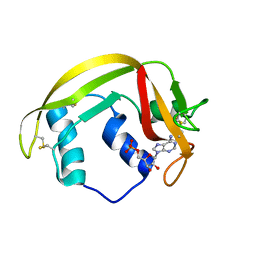
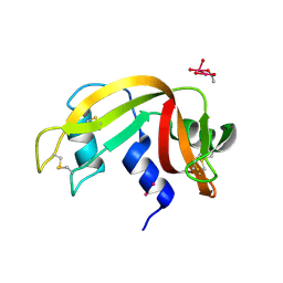
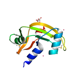
7TY1
 | | Crystal structure of apo eosinophil cationic protein (ribonuclease 3) from Macaca fascicularis (MfECP) | | 分子名称: | CITRIC ACID, DI(HYDROXYETHYL)ETHER, Eosinophil cationic protein, ... | | 著者 | Tran, T.T.Q, Pham, N.T.H, Calmettes, C, Doucet, N. | | 登録日 | 2022-02-11 | | 公開日 | 2023-08-16 | | 実験手法 | X-RAY DIFFRACTION (1.8 Å) | | 主引用文献 | Crystal structure of apo eosinophil cationic protein (ribonuclease 3) from Macaca fascicularis (MfECP)
To Be Published
|
|
9RAT
 | |
9RSA
 | |
7BFL
 | |
7BFK
 | | X-ray structure of SS-RNase-2 | | 分子名称: | Angiogenin-1 | | 著者 | Sica, F, Russo Krauss, I, Troisi, R. | | 登録日 | 2021-01-04 | | 公開日 | 2021-04-28 | | 最終更新日 | 2024-01-31 | | 実験手法 | X-RAY DIFFRACTION (1.89 Å) | | 主引用文献 | The structural features of an ancient ribonuclease from Salmo salar reveal an intriguing case of auto-inhibition.
Int.J.Biol.Macromol., 182, 2021
|
|
6XW0
 | |
6XVX
 | |
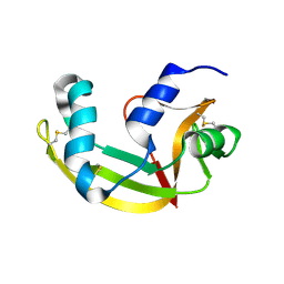
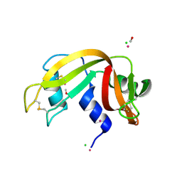
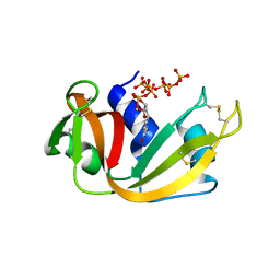
6MV6
 | | Crystal structure of RNAse 6 | | 分子名称: | PHOSPHATE ION, Ribonuclease K6 | | 著者 | Couture, J.-F, Doucet, N. | | 登録日 | 2018-10-24 | | 公開日 | 2019-11-13 | | 最終更新日 | 2020-05-27 | | 実験手法 | X-RAY DIFFRACTION (1.5 Å) | | 主引用文献 | Insights into Structural and Dynamical Changes Experienced by Human RNase 6 upon Ligand Binding.
Biochemistry, 59, 2020
|
|
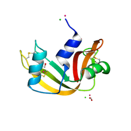
6MV7
 | | Crystal structure of RNAse 6 | | 分子名称: | ADENOSINE MONOPHOSPHATE, Ribonuclease K6 | | 著者 | Couture, J.-F, Doucet, N. | | 登録日 | 2018-10-24 | | 公開日 | 2019-11-13 | | 最終更新日 | 2023-10-11 | | 実験手法 | X-RAY DIFFRACTION (2.59 Å) | | 主引用文献 | Insights into Structural and Dynamical Changes Experienced by Human RNase 6 upon Ligand Binding.
Biochemistry, 59, 2020
|
|
5JMG
 | | X-ray structure of the complex between bovine pancreatic ribonuclease and pentachlorocarbonyliridate(III) (4 days of soaking) | | 分子名称: | CARBON MONOXIDE, CHLORIDE ION, IRIDIUM ION, ... | | 著者 | Caterino, M, Petruk, A.A, Vergara, A, Ferraro, G, Merlino, A. | | 登録日 | 2016-04-29 | | 公開日 | 2016-07-27 | | 最終更新日 | 2024-01-10 | | 実験手法 | X-RAY DIFFRACTION (1.85 Å) | | 主引用文献 | Mapping the protein-binding sites for iridium(iii)-based CO-releasing molecules.
Dalton Trans, 45, 2016
|
|
5JML
 | | X-ray structure of the complex between bovine pancreatic ribonuclease and penthachlorocarbonyliridate(III) (2 months of soaking) | | 分子名称: | CARBON MONOXIDE, CHLORIDE ION, IRIDIUM ION, ... | | 著者 | Caterino, M, Petruk, A.A, Vergara, A, Ferraro, G, Merlino, A. | | 登録日 | 2016-04-29 | | 公開日 | 2016-07-27 | | 最終更新日 | 2024-01-10 | | 実験手法 | X-RAY DIFFRACTION (2.29 Å) | | 主引用文献 | Mapping the protein-binding sites for iridium(iii)-based CO-releasing molecules.
Dalton Trans, 45, 2016
|
|
5JLG
 | | The X-ray structure of the adduct formed in the reaction between bovine pancreatic ribonuclease and compound I, a piano-stool organometallic Ru(II) arene compound containing an O,S-chelating ligand | | 分子名称: | DIMETHYL SULFOXIDE, RUTHENIUM ION, Ribonuclease pancreatic, ... | | 著者 | Ferraro, G, Merlino, A. | | 登録日 | 2016-04-27 | | 公開日 | 2016-08-03 | | 最終更新日 | 2024-01-10 | | 実験手法 | X-RAY DIFFRACTION (1.79 Å) | | 主引用文献 | Unusual mode of protein binding by a cytotoxic pi-arene ruthenium(ii) piano-stool compound containing an O,S-chelating ligand.
Dalton Trans, 45, 2016
|
|
7Z6D
 | |
7Z6G
 | |
8AF0
 | |
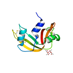
8F5X
 | | Crystal structure of human eosinophil-derived neurotoxin (EDN, ribonuclease 2) in complex with 5'-adenosine monophosphate (AMP) | | 分子名称: | 1,2-ETHANEDIOL, ADENOSINE MONOPHOSPHATE, Non-secretory ribonuclease, ... | | 著者 | Tran, T.T.Q, Pham, N.T.H, Calmettes, C, Doucet, N. | | 登録日 | 2022-11-15 | | 公開日 | 2023-11-29 | | 実験手法 | X-RAY DIFFRACTION (1.7 Å) | | 主引用文献 | Crystal structure of human eosinophil-derived neurotoxin (EDN, ribonuclease 2) in complex with 5'-adenosine monophosphate (AMP)
To Be Published
|
|
8FHM
 | | RNase A-Uridine 5'-Hexaphosphate (RNaseA.p6U) | | 分子名称: | 5'-O-[(S)-hydroxy{[(S)-hydroxy{[(R)-hydroxy{[(S)-hydroxy{[(R)-hydroxy(phosphonooxy)phosphoryl]oxy}phosphoryl]oxy}phosphoryl]oxy}phosphoryl]oxy}phosphoryl]uridine, Ribonuclease pancreatic | | 著者 | Park, G, Cummins, C. | | 登録日 | 2022-12-14 | | 公開日 | 2023-12-20 | | 実験手法 | SOLUTION SCATTERING (1.79 Å), X-RAY DIFFRACTION | | 主引用文献 | Experimental and Computational Studies for the Effect of Lengthening the Phosphate Chain of Nucleotide on the RNase A inhibitors.
To Be Published
|
|
7NPM
 | |
8GC9
 | | RNase A-Uridine 5'-Heptaphosphate (RNase A.p7U) | | 分子名称: | Ribonuclease pancreatic, uridine 5'-heptaphosphate | | 著者 | Park, G, Cummins, C. | | 登録日 | 2023-03-01 | | 公開日 | 2024-03-06 | | 実験手法 | X-RAY DIFFRACTION (1.85 Å) | | 主引用文献 | Experimental and Computational Studies for the Effect of Lengthening the Phosphate Chain of Nucleotide on the RNase A Inhibitors.
To Be Published
|
|
8GGG
 | | RNase A-Adenosine 5'-Hexaphosphate (RNaseA.p6A) | | 分子名称: | GLYCEROL, Ribonuclease pancreatic, adenosine 5'-hexaphosphate | | 著者 | Park, G, Cummins, C. | | 登録日 | 2023-03-08 | | 公開日 | 2024-03-13 | | 実験手法 | X-RAY DIFFRACTION (1.86 Å) | | 主引用文献 | Experimental and Computational Studies for the Effect of Lengthening the Phosphate Chain of Nucleotide on the RNase A Inhibitiors.
To Be Published
|
|
7OR6
 | |
7ORD
 | |
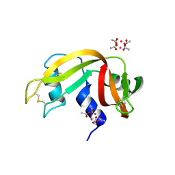
6PVU
 | | RNase A in complex with hexametaphosphate | | 分子名称: | 2,4,6,8,10,12-hexahydroxy-2lambda~5~,4lambda~5~,6lambda~5~,8lambda~5~,10lambda~5~,12lambda~5~-cyclohexaphosphoxane-2,4,6,8,10,12-hexone, Ribonuclease pancreatic | | 著者 | Windsor, I.W, Sheppard, S.M, Cummins, C.C, Raines, R.T. | | 登録日 | 2019-07-21 | | 公開日 | 2019-11-06 | | 最終更新日 | 2023-10-11 | | 実験手法 | X-RAY DIFFRACTION (1.49 Å) | | 主引用文献 | Nucleoside Tetra- and Pentaphosphates Prepared Using a Tetraphosphorylation Reagent Are Potent Inhibitors of Ribonuclease A.
J.Am.Chem.Soc., 141, 2019
|
|


