 Movie
Movie Controller


+ Open data

- Basic information
| Entry | Database: PDB / ID: 1tub | ||||||
|---|---|---|---|---|---|---|---|
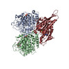
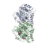
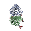
| Title | TUBULIN ALPHA-BETA DIMER, ELECTRON DIFFRACTION | ||||||
 Components Components | (TUBULIN ) x 2 ) x 2 | ||||||
 Keywords Keywords | MICROTUBULES / ALPHA-TUBULIN / BETA-TUBULIN / GTPASE | ||||||
| Function / homology |  Function and homology informationHydrolases; Acting on acid anhydrides; Acting on GTP to facilitate cellular and subcellular movement / structural constituent of cytoskeleton / microtubule cytoskeleton organization / mitotic cell cycle / microtubule / hydrolase activity / GTPase activity / GTP binding / metal ion binding / cytoplasm Function and homology informationHydrolases; Acting on acid anhydrides; Acting on GTP to facilitate cellular and subcellular movement / structural constituent of cytoskeleton / microtubule cytoskeleton organization / mitotic cell cycle / microtubule / hydrolase activity / GTPase activity / GTP binding / metal ion binding / cytoplasmSimilarity search - Function | ||||||
| Biological species |  Sus scrofa (pig) Sus scrofa (pig) | ||||||
| Method | ELECTRON CRYSTALLOGRAPHY / electron crystallography / cryo EM / Resolution: 3.7 Å | ||||||
 Authors Authors | Nogales, E. / Downing, K.H. | ||||||
 Citation Citation | Journal: Nature / Year: 1998 Title: Structure of the alpha beta tubulin dimer by electron crystallography. Authors: E Nogales / S G Wolf / K H Downing /  Abstract: The alphabeta tubulin heterodimer is the structural subunit of microtubules, which are cytoskeletal elements that are essential for intracellular transport and cell division in all eukaryotes. Each ...The alphabeta tubulin heterodimer is the structural subunit of microtubules, which are cytoskeletal elements that are essential for intracellular transport and cell division in all eukaryotes. Each tubulin monomer binds a guanine nucleotide, which is nonexchangeable when it is bound in the alpha subunit, or N site, and exchangeable when bound in the beta subunit, or E site. The alpha- and beta-tubulins share 40% amino-acid sequence identity, both exist in several isotype forms, and both undergo a variety of posttranslational modifications. Limited sequence homology has been found with the proteins FtsZ and Misato, which are involved in cell division in bacteria and Drosophila, respectively. Here we present an atomic model of the alphabeta tubulin dimer fitted to a 3.7-A density map obtained by electron crystallography of zinc-induced tubulin sheets. The structures of alpha- and beta-tubulin are basically identical: each monomer is formed by a core of two beta-sheets surrounded by alpha-helices. The monomer structure is very compact, but can be divided into three functional domains: the amino-terminal domain containing the nucleotide-binding region, an intermediate domain containing the Taxol-binding site, and the carboxy-terminal domain, which probably constitutes the binding surface for motor proteins. #1: Journal: Nature / Year: 1998Title: Erratum. Structure of the Alpha Beta Tubulin Dimer by Electron Crystallography Authors: Nogales, E. / Wolf, S.G. / Downing, K.H. #2: Journal: Nat.Struct.Biol. / Year: 1998Title: Tubulin and Ftsz Form a Distinct Family of Gtpases Authors: Nogales, E. / Downing, K.H. / Amos, L.A. / Lowe, J. | ||||||
| History |
|
- Structure visualization
| Movie |
Movie viewer |
|---|---|
| Structure viewer | Molecule: MolmilJmol/JSmol |
- Downloads & links
-Download
| PDBx/mmCIF format | 1tub.cif.gz | 176.9 KB | Display | PDBx/mmCIF format |
|---|---|---|---|---|
| PDB format | pdb1tub.ent.gz | 128.8 KB | Display | PDB format |
| PDBx/mmJSON format | 1tub.json.gz | Tree view | PDBx/mmJSON format | |
| Others |  Other downloads Other downloads |
-Validation report
| Arichive directory | https://data.pdbj.org/pub/pdb/validation_reports/tu/1tubftp://data.pdbj.org/pub/pdb/validation_reports/tu/1tub | HTTPS FTP |
|---|
-Related structure data
| Related structure data | |
|---|---|
| Similar structure data |


-Links
 PDBj
PDBj


- Assembly
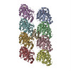
| Deposited unit | 
| ||||||||
|---|---|---|---|---|---|---|---|---|---|
| 1 |
| ||||||||
| Unit cell |
|
-Components
| #1: Protein | Mass: 48869.117 Da / Num. of mol.: 1 / Source method: isolated from a natural source / Source: (natural) Sus scrofa (pig) / Organ: BRAIN / References: UniProt: P02550 |
|---|---|
| #2: Protein | Mass: 47940.945 Da / Num. of mol.: 1 / Source method: isolated from a natural source / Source: (natural) Sus scrofa (pig) / Organ: BRAIN / References: UniProt: P02554 |
| #3: Chemical | ChemComp-GTP / Guanosine triphosphate  Mass: 523.180 Da / Num. of mol.: 1 / Source method: obtained synthetically / Formula: C10H16N5O14P3 / Comment: GTP, energy-carrying molecule*YM Mass: 523.180 Da / Num. of mol.: 1 / Source method: obtained synthetically / Formula: C10H16N5O14P3 / Comment: GTP, energy-carrying molecule*YM |
| #4: Chemical | ChemComp-GDP / Guanosine diphosphate  Type: RNA linking / Mass: 443.201 Da / Num. of mol.: 1 / Source method: obtained synthetically / Formula: C10H15N5O11P2 / Comment: GDP, energy-carrying molecule*YM Type: RNA linking / Mass: 443.201 Da / Num. of mol.: 1 / Source method: obtained synthetically / Formula: C10H15N5O11P2 / Comment: GDP, energy-carrying molecule*YM |
| #5: Chemical | ChemComp-TXL / Docetaxel  Mass: 807.879 Da / Num. of mol.: 1 / Source method: obtained synthetically / Formula: C43H53NO14 / Comment: medication, chemotherapy*YM Mass: 807.879 Da / Num. of mol.: 1 / Source method: obtained synthetically / Formula: C43H53NO14 / Comment: medication, chemotherapy*YM |
-Experimental details
-Experiment
| Experiment | Method: ELECTRON CRYSTALLOGRAPHY |
|---|---|
| EM experiment | Aggregation state: 2D ARRAY / 3D reconstruction method: electron crystallography |
- Sample preparation
| Component | Name: alpha-beta tubulin sheets / Type: COMPLEX |
|---|---|
| Specimen | Embedding applied: YES / Shadowing applied: NO / Staining applied: NO / Vitrification applied: YES |
| EM embedding | Material: tannin-glucose |
| Crystal | Description: THE MODEL WAS DERIVED USING ELECTRON DIFFRACTION AND IMAGE DATA FROM TWO DIMENSIONAL CRYSTALS OF TUBULIN INDUCED BY THE PRESENCE OF ZN++ IONS. WHAT FOLLOWS ARE THE COORDINATES FOR THE AB- ...Description: THE MODEL WAS DERIVED USING ELECTRON DIFFRACTION AND IMAGE DATA FROM TWO DIMENSIONAL CRYSTALS OF TUBULIN INDUCED BY THE PRESENCE OF ZN++ IONS. WHAT FOLLOWS ARE THE COORDINATES FOR THE AB-TUBULIN DIMER BOUND TO TAXOL AS OBTAINED BY ELECTRON CRYSTALLOGRAPHY OF ZINC-INDUCED SHEETS. THIS IS THE UNREFINED MODEL, BUILT INTO A RAW DENSITY MAP WHERE THE RESOLUTION IN THE PLANE OF THE SHEET WAS 3.7 ANGSTROMS AND THAT PERPENDICULAR TO THE SHEET ABOUT 4.8 ANGSTROMS. THE MODEL DOES NOT CONTAIN MOST OF THE C-TERMINAL RESIDUES OF EITHER MONOMER WHICH WERE DISORDERED IN THE MAP. THE LOOP BETWEEN HELIX H1 AND STRAND S2, AND THAT BETWEEN H2 AND S3 ARE PRESENT FOR COMPLETENESS BUT WERE BUILT INTO VERY WEAK DENSITY. GIVEN THE LIMITED RESOLUTION OF THE MAP, THE CONFORMATION OF THE SIDE CHAINS, ESPECIALLY THOSE CORRESPONDING TO RESIDUES ON THE SURFACE OF THE DIMER, MUST BE TAKEN CAUTIOUSLY. IN ADDITION, BECAUSE THIS IS AN UNREFINED MODEL, CERTAIN GEOMETRY ERRORS MAY STILL BE PRESENT IN THE STRUCTURE. PLEASE TAKE THIS INTO ACCOUNT WHEN INTERPRETING YOUR OWN DATA BASED ON THE PRESENT TUBULIN STRUCTURE. ALTHOUGH THE POSITION OF RESIDUES (WITH THE EXCEPTION OF THOSE IN THE LOOPS MENTIONED ABOVE) SHOULD NOT CHANGE SIGNIFICANTLY UPON REFINEMENT, DRAWING INFORMATION AT THE LEVEL OF SIDE CHAIN CONFORMATION IS CLEARLY NOT ADVISED. FINALLY, PLEASE NOTICE THAT THE TAXOID IN THE MODEL IS THE TAXOL DERIVATIVE TAXOTERE. |
| Crystal grow | Details: TWO-DIMENSIONAL SHEETS ARE FORMED UNDER NORMAL POLYMERIZING CONDITIONS WITH THE ADDITION OF 1 MM ZNCL2 |
| Crystal grow | *PLUS Method: unknown |
-Data collection
| EM imaging |
| |||||||||||||||||||||
|---|---|---|---|---|---|---|---|---|---|---|---|---|---|---|---|---|---|---|---|---|---|---|
| Image recording | Film or detector model: GENERIC FILM | |||||||||||||||||||||
| Detector | Date: Sep 1, 1996 | |||||||||||||||||||||
| Radiation wavelength | Relative weight: 1 | |||||||||||||||||||||
| Reflection | *PLUS Rmerge(I) obs: 0.25 |
- Processing
| Refinement | Highest resolution: 3.7 Å Details: THESE INITIAL COORDINATES ARE FOR THE MODEL BUILT IN THE ORIGINAL DENSITY MAP, WITH NO REFINEMENT. | ||||||||||||
|---|---|---|---|---|---|---|---|---|---|---|---|---|---|
| Refinement step | Cycle: LAST / Highest resolution: 3.7 Å
|
