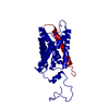
gap junction-mediated intercellular transport / water channel activity / water transport / structural constituent of eye lens / gap junction / response to stimulus / lens development in camera-type eye / positive regulation of cell adhesion / visual perception / protein homotetramerization ...gap junction-mediated intercellular transport / water channel activity / water transport / structural constituent of eye lens / gap junction / response to stimulus / lens development in camera-type eye / positive regulation of cell adhesion / visual perception / protein homotetramerization / calmodulin binding / endoplasmic reticulum / plasma membrane Similarity search - Function
Glycerol uptake facilitator protein / Glycerol uptake facilitator protein. / Aquaporin transporter / Major intrinsic protein, conserved site / MIP family signature. / Major intrinsic protein / Major intrinsic protein / Aquaporin-like / Up-down Bundle / Mainly Alpha Similarity search - Domain/homology
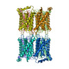
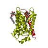
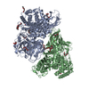
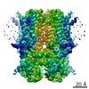
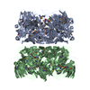
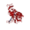
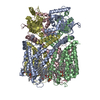
Journal: Nature / Year: 2005 Title: Lipid-protein interactions in double-layered two-dimensional AQP0 crystals. Authors: Tamir Gonen / Yifan Cheng / Piotr Sliz / Yoko Hiroaki / Yoshinori Fujiyoshi / Stephen C Harrison / Thomas Walz / Abstract: Lens-specific aquaporin-0 (AQP0) functions as a specific water pore and forms the thin junctions between fibre cells. Here we describe a 1.9 A resolution structure of junctional AQP0, determined by ...Lens-specific aquaporin-0 (AQP0) functions as a specific water pore and forms the thin junctions between fibre cells. Here we describe a 1.9 A resolution structure of junctional AQP0, determined by electron crystallography of double-layered two-dimensional crystals. Comparison of junctional and non-junctional AQP0 structures shows that junction formation depends on a conformational switch in an extracellular loop, which may result from cleavage of the cytoplasmic amino and carboxy termini. In the centre of the water pathway, the closed pore in junctional AQP0 retains only three water molecules, which are too widely spaced to form hydrogen bonds with each other. Packing interactions between AQP0 tetramers in the crystalline array are mediated by lipid molecules, which assume preferred conformations. We were therefore able to build an atomic model for the lipid bilayer surrounding the AQP0 tetramers, and we describe lipid-protein interactions.
History
Deposition
Oct 3, 2005
Deposition site: RCSB / Processing site: RCSB
Revision 1.0
Dec 6, 2005
Provider: repository / Type: Initial release
Revision 1.1
May 1, 2008
Group: Version format compliance
Revision 1.2
Jul 13, 2011
Group: Derived calculations / Version format compliance
EXPERIMENT TYPE : ELECTRON DIFFRACTION DATE OF DATA COLLECTION : 01-DEC-03 TEMPERATURE (KELVIN) : ...EXPERIMENT TYPE : ELECTRON DIFFRACTION DATE OF DATA COLLECTION : 01-DEC-03 TEMPERATURE (KELVIN) : 300.0 PH : 6.00 NUMBER OF CRYSTALS USED : 286 RADIATION SOURCE : JEM3000SFF OPTICS : CRYSTALS TILTED TO MAX : 71.3 DEGREES DETECTOR TYPE : CCD DETECTOR MANUFACTURER : GATAN 2K X 2K DATA SCALING SOFTWARE : GATAN ACCELERATION VOLTAGE (KV) : 300 NUMBER OF UNIQUE REFLECTIONS : 22293 RESOLUTION RANGE HIGH (A) : 1.9 RESOLUTION RANGE LOW (A) : 20.000 OVERALL. COMPLETENESS FOR RANGE (%) : 80.0 DATA REDUNDANCY : 5.700 IN THE HIGHEST RESOLUTION SHELL. HIGHEST RESOLUTION SHELL, RANGE HIGH (A) : 1.90 HIGHEST RESOLUTION SHELL, RANGE LOW (A) : 2.0 COMPLETENESS FOR SHELL (%) : 82.0 DATA REDUNDANCY IN SHELL : 5.70 R MERGE FOR SHELL (I) : 0.166 METHOD USED TO DETERMINE THE STRUCTURE: MOLECULAR : REPLACEMENT SOFTWARE USED: CNS STARTING MODEL: PDB ENTRY 1SOR
-
Structure visualization
Movie
Biological unit as author_and_software_defined_assembly
In the structure databanks used in Yorodumi, some data are registered as the other names, "COVID-19 virus" and "2019-nCoV". Here are the details of the virus and the list of structure data.
Jan 31, 2019. EMDB accession codes are about to change! (news from PDBe EMDB page)
EMDB accession codes are about to change! (news from PDBe EMDB page)
The allocation of 4 digits for EMDB accession codes will soon come to an end. Whilst these codes will remain in use, new EMDB accession codes will include an additional digit and will expand incrementally as the available range of codes is exhausted. The current 4-digit format prefixed with “EMD-” (i.e. EMD-XXXX) will advance to a 5-digit format (i.e. EMD-XXXXX), and so on. It is currently estimated that the 4-digit codes will be depleted around Spring 2019, at which point the 5-digit format will come into force.
The EM Navigator/Yorodumi systems omit the EMD- prefix.
Related info.:Q: What is EMD? / ID/Accession-code notation in Yorodumi/EM Navigator
Yorodumi is a browser for structure data from EMDB, PDB, SASBDB, etc.
This page is also the successor to EM Navigator detail page, and also detail information page/front-end page for Omokage search.
The word "yorodu" (or yorozu) is an old Japanese word meaning "ten thousand". "mi" (miru) is to see.
Related info.:EMDB / PDB / SASBDB / Comparison of 3 databanks / Yorodumi Search / Aug 31, 2016. New EM Navigator & Yorodumi / Yorodumi Papers / Jmol/JSmol / Function and homology information / Changes in new EM Navigator and Yorodumi
 Movie
Movie Controller

 Yorodumi
 Open data
 Basic information
 Components
 Keywords
 Function and homology information
 Authors
 Citation
 Structure visualization
 Downloads & links
 Other downloads
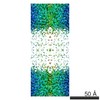

 PDBj
PDBj
 Assembly

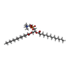
 Mass: 677.933 Da / Num. of mol.: 9 / Source method: obtained synthetically / Formula: C36H72NO8P / Comment: phospholipid*YM
 Mass: 18.015 Da / Num. of mol.: 79 / Source method: isolated from a natural source / Formula: H2O
 Sample preparation
 Processing