 Movie
Movie Controller

[English] 日本語
 Yorodumi
- PDB-5sy1: Structure of the STRA6 receptor for retinol uptake in complex wit... -
+ Open data

- Basic information
| Entry | Database: PDB / ID: 5sy1 | ||||||
|---|---|---|---|---|---|---|---|
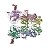
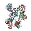
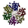
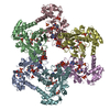
| Title | Structure of the STRA6 receptor for retinol uptake in complex with calmodulin | ||||||
 Components Components |
| ||||||
 Keywords Keywords | MEMBRANE PROTEIN/CALCIUM BINDING PROTEIN / Vitamin A / retinol / STRA6 / membrane / MEMBRANE PROTEIN-CALCIUM BINDING PROTEIN complex | ||||||
| Function / homology |  Function and homology information Function and homology informationvitamin A import into cell / The canonical retinoid cycle in rods (twilight vision) / retinol transport / retinol transmembrane transporter activity / chordate embryonic development / retinal binding / retinol binding / plasma membrane => GO:0005886 / calcium-mediated signaling / signaling receptor activity ...vitamin A import into cell / The canonical retinoid cycle in rods (twilight vision) / retinol transport / retinol transmembrane transporter activity / chordate embryonic development / retinal binding / retinol binding / plasma membrane => GO:0005886 / calcium-mediated signaling / signaling receptor activity / molecular adaptor activity / calmodulin binding / calcium ion binding / identical protein binding / plasma membrane Similarity search - Function | ||||||
| Biological species |    Spodoptera frugiperda (fall armyworm) Spodoptera frugiperda (fall armyworm) | ||||||
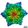
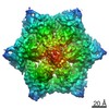
| Method | ELECTRON MICROSCOPY / single particle reconstruction / cryo EM / Resolution: 3.9 Å | ||||||
 Authors Authors | Clarke, O.B. / Chen, Y. / Mancia, F. | ||||||
 Citation Citation | Journal: Science / Year: 2016 Title: Structure of the STRA6 receptor for retinol uptake. Authors: Yunting Chen / Oliver B Clarke / Jonathan Kim / Sean Stowe / Youn-Kyung Kim / Zahra Assur / Michael Cavalier / Raquel Godoy-Ruiz / Desiree C von Alpen / Chiara Manzini / William S Blaner / ...Authors: Yunting Chen / Oliver B Clarke / Jonathan Kim / Sean Stowe / Youn-Kyung Kim / Zahra Assur / Michael Cavalier / Raquel Godoy-Ruiz / Desiree C von Alpen / Chiara Manzini / William S Blaner / Joachim Frank / Loredana Quadro / David J Weber / Lawrence Shapiro / Wayne A Hendrickson / Filippo Mancia /  Abstract: Vitamin A homeostasis is critical to normal cellular function. Retinol-binding protein (RBP) is the sole specific carrier in the bloodstream for hydrophobic retinol, the main form in which vitamin A ...Vitamin A homeostasis is critical to normal cellular function. Retinol-binding protein (RBP) is the sole specific carrier in the bloodstream for hydrophobic retinol, the main form in which vitamin A is transported. The integral membrane receptor STRA6 mediates cellular uptake of vitamin A by recognizing RBP-retinol to trigger release and internalization of retinol. We present the structure of zebrafish STRA6 determined to 3.9-angstrom resolution by single-particle cryo-electron microscopy. STRA6 has one intramembrane and nine transmembrane helices in an intricate dimeric assembly. Unexpectedly, calmodulin is bound tightly to STRA6 in a noncanonical arrangement. Residues involved with RBP binding map to an archlike structure that covers a deep lipophilic cleft. This cleft is open to the membrane, suggesting a possible mode for internalization of retinol through direct diffusion into the lipid bilayer. | ||||||
| History |
|
- Structure visualization
| Movie |
Movie viewer |
|---|---|
| Structure viewer | Molecule: MolmilJmol/JSmol |
- Downloads & links
-Download
| PDBx/mmCIF format | 5sy1.cif.gz | 271.8 KB | Display | PDBx/mmCIF format |
|---|---|---|---|---|
| PDB format | pdb5sy1.ent.gz | 218.3 KB | Display | PDB format |
| PDBx/mmJSON format | 5sy1.json.gz | Tree view | PDBx/mmJSON format | |
| Others |  Other downloads Other downloads |
-Validation report
| Summary document | 5sy1_validation.pdf.gz | 1.5 MB | Display | wwPDB validaton report |
|---|---|---|---|---|
| Full document | 5sy1_full_validation.pdf.gz | 1.5 MB | Display | |
| Data in XML | 5sy1_validation.xml.gz | 49.4 KB | Display | |
| Data in CIF | 5sy1_validation.cif.gz | 75.3 KB | Display | |
| Arichive directory | https://data.pdbj.org/pub/pdb/validation_reports/sy/5sy1ftp://data.pdbj.org/pub/pdb/validation_reports/sy/5sy1 | HTTPS FTP |
-Related structure data
| Related structure data | 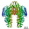 8315MC  5k8qC M: map data used to model this data C: citing same article ( |
|---|---|
| Similar structure data |


-Links
 PDBj
PDBj


- Assembly
| Deposited unit | 
|
|---|---|
| 1 |
|
-Components
| #1: Protein | Mass: 16825.520 Da / Num. of mol.: 2 / Source method: isolated from a natural source / Source: (natural) Spodoptera frugiperda (fall armyworm) / References: UniProt: A0A1C7D1B9*PLUS#2: Protein | Mass: 75551.523 Da / Num. of mol.: 2 Source method: isolated from a genetically manipulated source Source: (gene. exp.) Spodoptera frugiperda (fall armyworm) / References: UniProt: A4IGB6, UniProt: F1RAX4*PLUS#3: Chemical | ChemComp-CA /   Mass: 40.078 Da / Num. of mol.: 8 / Source method: obtained synthetically / Formula: Ca Mass: 40.078 Da / Num. of mol.: 8 / Source method: obtained synthetically / Formula: Ca#4: Chemical |   Mass: 386.654 Da / Num. of mol.: 2 / Source method: obtained synthetically / Formula: C27H46O Mass: 386.654 Da / Num. of mol.: 2 / Source method: obtained synthetically / Formula: C27H46O |
|---|
-Experimental details
-Experiment
| Experiment | Method: ELECTRON MICROSCOPY |
|---|---|
| EM experiment | Aggregation state: PARTICLE / 3D reconstruction method: single particle reconstruction |
- Sample preparation
| Component | Name: Complex of zebrafish (D. rerio) STRA6 with copurified calmodulin reconstituted in amphipol A8-35 Type: COMPLEX / Entity ID: #1-#2 / Source: RECOMBINANT |
|---|---|
| Source (natural) | Organism: |
| Source (recombinant) | Organism: Spodoptera frugiperda (fall armyworm) / Plasmid: pIEX/Bac-1 |
| Buffer solution | pH: 7 |
| Specimen | Conc.: 0.6 mg/ml / Embedding applied: NO / Shadowing applied: NO / Staining applied: NO / Vitrification applied: YES |
| Specimen support | Grid material: GOLD / Grid mesh size: 400 divisions/in. / Grid type: Quantifoil, UltrAuFoil, R1.2/1.3 |
| Vitrification | Instrument: FEI VITROBOT MARK IV / Cryogen name: ETHANE / Humidity: 95 % / Chamber temperature: 277 K Details: 3 uL sample applied, 3-4 second blot time, 30 second wait time, blot force 3, grid blotted from both sides, plunged into liquid ethane (FEI VITROBOT MARK IV). |
- Electron microscopy imaging
| Experimental equipment |  Model: Tecnai F30 / Image courtesy: FEI Company |
|---|---|
| Microscopy | Model: FEI TECNAI F30 |
| Electron gun | Electron source:  FIELD EMISSION GUN / Accelerating voltage: 300 kV / Illumination mode: FLOOD BEAM FIELD EMISSION GUN / Accelerating voltage: 300 kV / Illumination mode: FLOOD BEAM |
| Electron lens | Mode: BRIGHT FIELD |
| Image recording | Electron dose: 100 e/Å2 / Detector mode: COUNTING / Film or detector model: GATAN K2 SUMMIT (4k x 4k) |
- Processing
| Software | Name: PHENIX / Version: dev_2427: / Classification: refinement | ||||||||||||||||||||||||||||||||||||||||
|---|---|---|---|---|---|---|---|---|---|---|---|---|---|---|---|---|---|---|---|---|---|---|---|---|---|---|---|---|---|---|---|---|---|---|---|---|---|---|---|---|---|
| EM software |
| ||||||||||||||||||||||||||||||||||||||||
| CTF correction | Type: PHASE FLIPPING AND AMPLITUDE CORRECTION | ||||||||||||||||||||||||||||||||||||||||
| Symmetry | Point symmetry: C2 (2 fold cyclic) | ||||||||||||||||||||||||||||||||||||||||
| 3D reconstruction | Resolution: 3.9 Å / Resolution method: FSC 0.143 CUT-OFF / Num. of particles: 56615 / Symmetry type: POINT | ||||||||||||||||||||||||||||||||||||||||
| Atomic model building | Protocol: AB INITIO MODEL | ||||||||||||||||||||||||||||||||||||||||
| Refine LS restraints |
|
