Movie
Movie Controller

[English] 日本語
 Yorodumi
- PDB-5jlh: Cryo-EM structure of a human cytoplasmic actomyosin complex at ne... -
+ Open data

- Basic information
| Entry | Database: PDB / ID: 5jlh | |||||||||
|---|---|---|---|---|---|---|---|---|---|---|
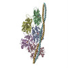
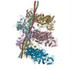
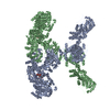
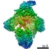
| Title | Cryo-EM structure of a human cytoplasmic actomyosin complex at near-atomic resolution | |||||||||
 Components Components |
| |||||||||
 Keywords Keywords |  CONTRACTILE PROTEIN / CONTRACTILE FILAMENT / MUSCLE / THIN FILAMENT / CYTOSKELETON / STRUCTURAL PROTEIN / HYDROLASE COMPLEX / F-actin / tropomyosin / filament / myosin / protein polymers / cryo EM CONTRACTILE PROTEIN / CONTRACTILE FILAMENT / MUSCLE / THIN FILAMENT / CYTOSKELETON / STRUCTURAL PROTEIN / HYDROLASE COMPLEX / F-actin / tropomyosin / filament / myosin / protein polymers / cryo EM | |||||||||
| Function / homology |  Function and homology informationcontractile vacuole / COPI-mediated anterograde transport / basal body patch / Platelet degranulation / sorocarp development / myosin II filament / tight junction assembly / Neutrophil degranulation / macropinocytic cup / actin filament-based movement ...contractile vacuole / COPI-mediated anterograde transport / basal body patch / Platelet degranulation / sorocarp development / myosin II filament / tight junction assembly / Neutrophil degranulation / macropinocytic cup / actin filament-based movement / actin crosslink formation / regulation of transepithelial transport / : / structural constituent of postsynaptic actin cytoskeleton / protein localization to bicellular tight junction / morphogenesis of a polarized epithelium / profilin binding / Formation of annular gap junctions / vocalization behavior / Gap junction degradation / dense body / Cell-extracellular matrix interactions / actomyosin / myosin filament / actomyosin structure organization / myosin II complex / RHO GTPases Activate ROCKs / regulation of stress fiber assembly / RHO GTPases activate CIT / Adherens junctions interactions / hyperosmotic response / Sema4D induced cell migration and growth-cone collapse / Sensory processing of sound by outer hair cells of the cochlea / Sensory processing of sound by inner hair cells of the cochlea / Interaction between L1 and Ankyrins / sarcomere organization / NuA4 histone acetyltransferase complex / regulation of synaptic vesicle endocytosis / microfilament motor activity / apical junction complex / regulation of focal adhesion assembly / maintenance of blood-brain barrier / cortical actin cytoskeleton / positive regulation of wound healing / myofibril / EPHA-mediated growth cone collapse / Recycling pathway of L1 / cell leading edge / filamentous actin / RHO GTPases activate PAKs / pseudopodium / actin filament bundle assembly / brush border / neuronal action potential / calyx of Held / skeletal muscle contraction / EPH-ephrin mediated repulsion of cells / RHO GTPases Activate WASPs and WAVEs / RHO GTPases activate IQGAPs / RHOBTB2 GTPase cycle / phagocytic vesicle / phagocytosis / skeletal muscle tissue development / stress fiber / RHO GTPases activate PKNs / EPHB-mediated forward signaling / cellular response to starvation / extracellular matrix / axonogenesis / cell projection / cell motility / actin filament / RHO GTPases Activate Formins / Translocation of SLC2A4 (GLUT4) to the plasma membrane / sensory perception of sound / FCGR3A-mediated phagocytosis / Hydrolases; Acting on acid anhydrides; Acting on acid anhydrides to facilitate cellular and subcellular movement / Signaling by high-kinase activity BRAF mutants / Schaffer collateral - CA1 synapse / MAP2K and MAPK activation / structural constituent of cytoskeleton / Regulation of actin dynamics for phagocytic cup formation / platelet aggregation / VEGFA-VEGFR2 Pathway / cellular response to type II interferon / Signaling by RAF1 mutants / Signaling by moderate kinase activity BRAF mutants / Paradoxical activation of RAF signaling by kinase inactive BRAF / Signaling downstream of RAS mutants / actin filament binding / cell-cell junction / Signaling by BRAF and RAF1 fusions / protein-macromolecule adaptor activity / cell junction / Clathrin-mediated endocytosis / cell cortex / regulation of cell shape / growth cone / actin cytoskeleton organization / angiogenesis Function and homology informationcontractile vacuole / COPI-mediated anterograde transport / basal body patch / Platelet degranulation / sorocarp development / myosin II filament / tight junction assembly / Neutrophil degranulation / macropinocytic cup / actin filament-based movement ...contractile vacuole / COPI-mediated anterograde transport / basal body patch / Platelet degranulation / sorocarp development / myosin II filament / tight junction assembly / Neutrophil degranulation / macropinocytic cup / actin filament-based movement / actin crosslink formation / regulation of transepithelial transport / : / structural constituent of postsynaptic actin cytoskeleton / protein localization to bicellular tight junction / morphogenesis of a polarized epithelium / profilin binding / Formation of annular gap junctions / vocalization behavior / Gap junction degradation / dense body / Cell-extracellular matrix interactions / actomyosin / myosin filament / actomyosin structure organization / myosin II complex / RHO GTPases Activate ROCKs / regulation of stress fiber assembly / RHO GTPases activate CIT / Adherens junctions interactions / hyperosmotic response / Sema4D induced cell migration and growth-cone collapse / Sensory processing of sound by outer hair cells of the cochlea / Sensory processing of sound by inner hair cells of the cochlea / Interaction between L1 and Ankyrins / sarcomere organization / NuA4 histone acetyltransferase complex / regulation of synaptic vesicle endocytosis / microfilament motor activity / apical junction complex / regulation of focal adhesion assembly / maintenance of blood-brain barrier / cortical actin cytoskeleton / positive regulation of wound healing / myofibril / EPHA-mediated growth cone collapse / Recycling pathway of L1 / cell leading edge / filamentous actin / RHO GTPases activate PAKs / pseudopodium / actin filament bundle assembly / brush border / neuronal action potential / calyx of Held / skeletal muscle contraction / EPH-ephrin mediated repulsion of cells / RHO GTPases Activate WASPs and WAVEs / RHO GTPases activate IQGAPs / RHOBTB2 GTPase cycle / phagocytic vesicle / phagocytosis / skeletal muscle tissue development / stress fiber / RHO GTPases activate PKNs / EPHB-mediated forward signaling / cellular response to starvation / extracellular matrix / axonogenesis / cell projection / cell motility / actin filament / RHO GTPases Activate Formins / Translocation of SLC2A4 (GLUT4) to the plasma membrane / sensory perception of sound / FCGR3A-mediated phagocytosis / Hydrolases; Acting on acid anhydrides; Acting on acid anhydrides to facilitate cellular and subcellular movement / Signaling by high-kinase activity BRAF mutants / Schaffer collateral - CA1 synapse / MAP2K and MAPK activation / structural constituent of cytoskeleton / Regulation of actin dynamics for phagocytic cup formation / platelet aggregation / VEGFA-VEGFR2 Pathway / cellular response to type II interferon / Signaling by RAF1 mutants / Signaling by moderate kinase activity BRAF mutants / Paradoxical activation of RAF signaling by kinase inactive BRAF / Signaling downstream of RAS mutants / actin filament binding / cell-cell junction / Signaling by BRAF and RAF1 fusions / protein-macromolecule adaptor activity / cell junction / Clathrin-mediated endocytosis / cell cortex / regulation of cell shape / growth cone / actin cytoskeleton organization / angiogenesisSimilarity search - Function | |||||||||
| Biological species |  Homo sapiens (human) Homo sapiens (human) Dictyostelium discoideum (eukaryote) Dictyostelium discoideum (eukaryote) | |||||||||
| Method | ELECTRON MICROSCOPY / single particle reconstruction / cryo EM / Resolution: 3.9 Å | |||||||||
 Authors Authors | von der Ecken, J. / Heissler, S.M. / Pathan-Chhatbar, S. / Manstein, D.J. / Raunser, S. | |||||||||
| Funding support |  Germany, 2items Germany, 2items
| |||||||||
 Citation Citation | Journal: Nature / Year: 2016 Title: Cryo-EM structure of a human cytoplasmic actomyosin complex at near-atomic resolution. Abstract: The interaction of myosin with actin filaments is the central feature of muscle contraction and cargo movement along actin filaments of the cytoskeleton. The energy for these movements is generated ...The interaction of myosin with actin filaments is the central feature of muscle contraction and cargo movement along actin filaments of the cytoskeleton. The energy for these movements is generated during a complex mechanochemical reaction cycle. Crystal structures of myosin in different states have provided important structural insights into the myosin motor cycle when myosin is detached from F-actin. The difficulty of obtaining diffracting crystals, however, has prevented structure determination by crystallography of actomyosin complexes. Thus, although structural models exist of F-actin in complex with various myosins, a high-resolution structure of the F-actin–myosin complex is missing. Here, using electron cryomicroscopy, we present the structure of a human rigor actomyosin complex at an average resolution of 3.9 Å. The structure reveals details of the actomyosin interface, which is mainly stabilized by hydrophobic interactions. The negatively charged amino (N) terminus of actin interacts with a conserved basic motif in loop 2 of myosin, promoting cleft closure in myosin. Surprisingly, the overall structure of myosin is similar to rigor-like myosin structures in the absence of F-actin, indicating that F-actin binding induces only minimal conformational changes in myosin. A comparison with pre-powerstroke and intermediate (Pi-release) states of myosin allows us to discuss the general mechanism of myosin binding to F-actin. Our results serve as a strong foundation for the molecular understanding of cytoskeletal diseases, such as autosomal dominant hearing loss and diseases affecting skeletal and cardiac muscles, in particular nemaline myopathy and hypertrophic cardiomyopathy. | |||||||||
| History |
|
- Structure visualization
| Movie |
Movie viewer |
|---|---|
| Structure viewer | Molecule: MolmilJmol/JSmol |
- Downloads & links
-Download
| PDBx/mmCIF format | 5jlh.cif.gz | 729 KB | Display | PDBx/mmCIF format |
|---|---|---|---|---|
| PDB format | pdb5jlh.ent.gz | 588.6 KB | Display | PDB format |
| PDBx/mmJSON format | 5jlh.json.gz | Tree view | PDBx/mmJSON format | |
| Others |  Other downloads Other downloads |
-Validation report
| Arichive directory | https://data.pdbj.org/pub/pdb/validation_reports/jl/5jlhftp://data.pdbj.org/pub/pdb/validation_reports/jl/5jlh | HTTPS FTP |
|---|
-Related structure data
| Related structure data | 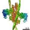 8164MC 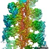 8165MC 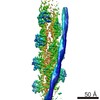 8162C  8163C 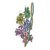 5jlfC M: map data used to model this data C: citing same article ( |
|---|---|
| Similar structure data |


-Links
 PDBj
PDBj


- Assembly
| Deposited unit | 
| |||||||||||||||||||||||||||||||||||||||||||||||||||||||||||||||||||||||||||||||||||||||||||
|---|---|---|---|---|---|---|---|---|---|---|---|---|---|---|---|---|---|---|---|---|---|---|---|---|---|---|---|---|---|---|---|---|---|---|---|---|---|---|---|---|---|---|---|---|---|---|---|---|---|---|---|---|---|---|---|---|---|---|---|---|---|---|---|---|---|---|---|---|---|---|---|---|---|---|---|---|---|---|---|---|---|---|---|---|---|---|---|---|---|---|---|---|
| 1 |
| |||||||||||||||||||||||||||||||||||||||||||||||||||||||||||||||||||||||||||||||||||||||||||
| Noncrystallographic symmetry (NCS) | NCS domain:
NCS domain segments:
NCS ensembles :
NCS oper:
|
-Components
| #1: Protein | / Gamma-actin Mass: 41707.570 Da / Num. of mol.: 5 Source method: isolated from a genetically manipulated source Source: (gene. exp.) Homo sapiens (human) / Gene: ACTG1, ACTG / Production host:   Spodoptera frugiperda (fall armyworm) / References: UniProt: P63261 Spodoptera frugiperda (fall armyworm) / References: UniProt: P63261#2: Protein | Mass: 117570.633 Da / Num. of mol.: 2 Source method: isolated from a genetically manipulated source Details: NON MUSCLE MYOSIN 2C HEAVY CHAIN, MOTOR DOMAIN RESIDUES 1-799 LINKED TO ALPHA-ACTININ 3, REPEATS 1 AND 2 RESIDUES 800-1039 FRAGMENT: UNP Q7Z406-1 RESIDUES 1-799, UNP P05095 RESIDUES 265-502 Source: (gene. exp.) Homo sapiens (human), (gene. exp.) Dictyostelium discoideum (eukaryote)Gene: MYH14, KIAA2034, FP17425, abpA, actnA, DDB_G0268632 / Production host: Spodoptera frugiperda (fall armyworm) / References: UniProt: Q7Z406, UniProt: P05095#3: Protein | Mass: 11507.176 Da / Num. of mol.: 4 Source method: isolated from a genetically manipulated source Details: Human TROPOMYSIN WAS USED (UNP P06753-2,TPM3_HUMAN, RESIDUES 62-196). DUE TO THE LIMITED RESOLUTION OF THE CRYO-EM DENSITY IN THE REGION OF TROPOMYOSIN, TROPOMYOSIN HAS BEEN REPRESENTED AS POLY(UNK). Source: (gene. exp.) Homo sapiens (human) / Production host:  Escherichia coli (E. coli) Escherichia coli (E. coli)#4: Chemical | ChemComp-ADP / Adenosine diphosphate  Mass: 427.201 Da / Num. of mol.: 5 / Source method: obtained synthetically / Formula: C10H15N5O10P2 / Comment: ADP, energy-carrying molecule*YM Mass: 427.201 Da / Num. of mol.: 5 / Source method: obtained synthetically / Formula: C10H15N5O10P2 / Comment: ADP, energy-carrying molecule*YM#5: Chemical | ChemComp-MG /   Mass: 24.305 Da / Num. of mol.: 5 / Source method: obtained synthetically / Formula: Mg Mass: 24.305 Da / Num. of mol.: 5 / Source method: obtained synthetically / Formula: MgSequence details | Human TROPOMYSIN WAS USED (UNP P06753-2,TPM3_HUMAN, RESIDUES 62-196) DUE TO THE LIMITED RESOLUTION ...Human TROPOMYSIN | |
|---|
-Experimental details
-Experiment
| Experiment | Method: ELECTRON MICROSCOPY |
|---|---|
| EM experiment | Aggregation state: FILAMENT / 3D reconstruction method: single particle reconstruction |
- Sample preparation
| Component |
| ||||||||||||||||||||||||||||
|---|---|---|---|---|---|---|---|---|---|---|---|---|---|---|---|---|---|---|---|---|---|---|---|---|---|---|---|---|---|
| Molecular weight | Value: 0.44 MDa / Experimental value: NO | ||||||||||||||||||||||||||||
| Source (natural) |
| ||||||||||||||||||||||||||||
| Source (recombinant) |
| ||||||||||||||||||||||||||||
| Buffer solution | pH: 7.5 Details: 5 mM Tris-HCl pH 7.5, 1 mM DTT, 100 mM KCl, and 2 mM MgCl2 | ||||||||||||||||||||||||||||
| Buffer component |
| ||||||||||||||||||||||||||||
| Specimen | Embedding applied: NO / Shadowing applied: NO / Staining applied: NO / Vitrification applied: YES | ||||||||||||||||||||||||||||
| Specimen support | Grid material: COPPER / Grid mesh size: 300 divisions/in. / Grid type: C-flat-2/1 | ||||||||||||||||||||||||||||
| EM embedding | Material: I | ||||||||||||||||||||||||||||
| Vitrification | Instrument: GATAN CRYOPLUNGE 3 / Cryogen name: ETHANE / Humidity: 90 % Details: Sample (2 uL of F-actin-tropomyosin solution) was applied to a glow-discharged holey carbon grid, incubated for 20 s and manually blotted from the backside for less than a second with filter ...Details: Sample (2 uL of F-actin-tropomyosin solution) was applied to a glow-discharged holey carbon grid, incubated for 20 s and manually blotted from the backside for less than a second with filter paper. Afterwards 1.5 uL of myosin solution (3 uM without nucleotide) were added directly on the grid, incubated for 10 s and then manually blotted for 5 s from the backside with filter paper. |
- Electron microscopy imaging
| Experimental equipment |  Model: Titan Krios / Image courtesy: FEI Company |
|---|---|
| Microscopy | Model: FEI TITAN KRIOS / Details: Cs corrected microscope |
| Electron gun | Electron source: FIELD EMISSION GUN / Accelerating voltage: 300 kV / Illumination mode: OTHER |
| Electron lens | Mode: BRIGHT FIELDBright-field microscopy / Nominal magnification: 59000 X / Calibrated defocus min: 700 nm / Calibrated defocus max: 2800 nm |
| Image recording | Average exposure time: 0.475 sec. / Electron dose: 16 e/Å2 / Detector mode: COUNTING / Film or detector model: FEI FALCON II (4k x 4k) / Num. of real images: 6300 |
| Image scans | Movie frames/image: 8 / Used frames/image: 2-8 |
- Processing
| Software | Name: REFMAC / Version: 5.8.0088 / Classification: refinement | ||||||||||||||||||||||||||||||||||||||||||||||||||||||||||||||||||||||||||||||||||||||||||||||||||||||||||
|---|---|---|---|---|---|---|---|---|---|---|---|---|---|---|---|---|---|---|---|---|---|---|---|---|---|---|---|---|---|---|---|---|---|---|---|---|---|---|---|---|---|---|---|---|---|---|---|---|---|---|---|---|---|---|---|---|---|---|---|---|---|---|---|---|---|---|---|---|---|---|---|---|---|---|---|---|---|---|---|---|---|---|---|---|---|---|---|---|---|---|---|---|---|---|---|---|---|---|---|---|---|---|---|---|---|---|---|
| EM software |
| ||||||||||||||||||||||||||||||||||||||||||||||||||||||||||||||||||||||||||||||||||||||||||||||||||||||||||
| CTF correction | Type: PHASE FLIPPING AND AMPLITUDE CORRECTION | ||||||||||||||||||||||||||||||||||||||||||||||||||||||||||||||||||||||||||||||||||||||||||||||||||||||||||
| Helical symmerty | Angular rotation/subunit: 166.9 ° / Axial rise/subunit: 27.5 Å / Axial symmetry: C1 | ||||||||||||||||||||||||||||||||||||||||||||||||||||||||||||||||||||||||||||||||||||||||||||||||||||||||||
| Particle selection | Num. of particles selected: 138000 | ||||||||||||||||||||||||||||||||||||||||||||||||||||||||||||||||||||||||||||||||||||||||||||||||||||||||||
| 3D reconstruction | Resolution: 3.9 Å / Resolution method: FSC 0.143 CUT-OFF / Num. of particles: 118000 Details: THE TROPOMYOSIN MAP FILTERED TO 7.0 ANGSTROM WAS MERGED WITH THE FINAL F-ACTIN-MYOSIN MAP (3.9 ANGSTROM) TO OBTAIN A MAP OF THE ENTIRE F-ACTIN-MYOSIN-TROPOMYOSIN COMPLEX. Symmetry type: HELICAL | ||||||||||||||||||||||||||||||||||||||||||||||||||||||||||||||||||||||||||||||||||||||||||||||||||||||||||
| Atomic model building |
| ||||||||||||||||||||||||||||||||||||||||||||||||||||||||||||||||||||||||||||||||||||||||||||||||||||||||||
| Atomic model building |
| ||||||||||||||||||||||||||||||||||||||||||||||||||||||||||||||||||||||||||||||||||||||||||||||||||||||||||
| Refinement | Resolution: 3.9→211.2 Å / Cor.coef. Fo:Fc: 0.907 / SU B: 27.204 / SU ML: 0.36 / ESU R: 0.558 Stereochemistry target values: MAXIMUM LIKELIHOOD WITH PHASES Details: HYDROGENS HAVE BEEN ADDED IN THE RIDING POSITIONS MERGED WITH THE FINAL F-ACTIN-MUYOSIN MAP (3.9 ANGSTROM) TO OBTAIN MAP OF THE ENTIRE F-ACTIN TROPOMYOSIN COMPLEX.
| ||||||||||||||||||||||||||||||||||||||||||||||||||||||||||||||||||||||||||||||||||||||||||||||||||||||||||
| Solvent computation | Ion probe radii: 0.8 Å / Shrinkage radii: 0.8 Å / VDW probe radii: 1.2 Å / Solvent model: MASK | ||||||||||||||||||||||||||||||||||||||||||||||||||||||||||||||||||||||||||||||||||||||||||||||||||||||||||
| Displacement parameters | Biso mean: 180.474 Å2
| ||||||||||||||||||||||||||||||||||||||||||||||||||||||||||||||||||||||||||||||||||||||||||||||||||||||||||
| Refinement step | Cycle: 1 / Total: 26477 | ||||||||||||||||||||||||||||||||||||||||||||||||||||||||||||||||||||||||||||||||||||||||||||||||||||||||||
| Refine LS restraints |
|