 Movie
Movie Controller


+ Open data

- Basic information
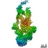
| Entry | Database: PDB / ID: 5afu | ||||||
|---|---|---|---|---|---|---|---|
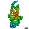
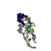
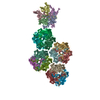
| Title | Cryo-EM structure of dynein tail-dynactin-BICD2N complex | ||||||
 Components Components |
| ||||||
 Keywords Keywords |  MOTOR PROTEIN / DYNEIN / DYNACTIN / BICD2 / MOTOR / TRANSPORT MOTOR PROTEIN / DYNEIN / DYNACTIN / BICD2 / MOTOR / TRANSPORT | ||||||
| Function / homology |  Function and homology information Function and homology informationretrograde axonal transport of mitochondrion / Gap junction degradation / Formation of annular gap junctions / Regulation of actin dynamics for phagocytic cup formation / EPHB-mediated forward signaling / VEGFA-VEGFR2 Pathway / Cell-extracellular matrix interactions / RHO GTPases Activate WASPs and WAVEs / MAP2K and MAPK activation / dynactin complex ...retrograde axonal transport of mitochondrion / Gap junction degradation / Formation of annular gap junctions / Regulation of actin dynamics for phagocytic cup formation / EPHB-mediated forward signaling / VEGFA-VEGFR2 Pathway / Cell-extracellular matrix interactions / RHO GTPases Activate WASPs and WAVEs / MAP2K and MAPK activation / dynactin complex / Clathrin-mediated endocytosis / F-actin capping protein complex / cellular response to cytochalasin B / regulation of transepithelial transport / structural constituent of postsynaptic actin cytoskeleton / morphogenesis of a polarized epithelium / postsynaptic actin cytoskeleton / protein localization to adherens junction / dense body / Tat protein binding / Neutrophil degranulation / dynein complex / apical protein localization / barbed-end actin filament capping / adherens junction assembly / coronary vasculature development / RHO GTPases activate IQGAPs / RHO GTPases Activate Formins / HSP90 chaperone cycle for steroid hormone receptors (SHR) in the presence of ligand / COPI-independent Golgi-to-ER retrograde traffic / MHC class II antigen presentation / tight junction / regulation of norepinephrine uptake / COPI-mediated anterograde transport / aorta development / NuA4 histone acetyltransferase complex / regulation of synaptic vesicle endocytosis / ventricular septum development / apical junction complex / establishment or maintenance of cell polarity / dynein complex binding / cortical cytoskeleton / positive regulation of double-strand break repair via homologous recombination / nitric-oxide synthase binding / brush border / kinesin binding / calyx of Held / regulation of protein localization to plasma membrane / microtubule-based process / axon cytoplasm / axonogenesis / mitotic spindle organization / actin filament / cell motility / adherens junction / Hydrolases; Acting on acid anhydrides; Acting on acid anhydrides to facilitate cellular and subcellular movement / Schaffer collateral - CA1 synapse / cytoplasmic ribonucleoprotein granule / kinetochore / nucleosome / actin cytoskeleton / lamellipodium / actin binding / cell cortex / actin cytoskeleton organization / nuclear membrane / cytoskeleton / regulation of cell cycle / hydrolase activity / ribonucleoprotein complex / axon / focal adhesion / centrosome / glutamatergic synapse / synapse / protein kinase binding / protein-containing complex / nucleoplasm / ATP binding / membrane / identical protein binding / nucleus / plasma membrane / cytosol / cytoplasmSimilarity search - Function | ||||||
| Biological species |  SUS SCROFA (pig) SUS SCROFA (pig) | ||||||
| Method | ELECTRON MICROSCOPY / single particle reconstruction / cryo EM / Resolution: 8.2 Å | ||||||
 Authors Authors | Urnavicius, L. / Zhang, K. / Diamant, A.G. / Motz, C. / Schlager, M.A. / Yu, M. / Patel, N.A. / Robinson, C.V. / Carter, A.P. | ||||||
 Citation Citation | Journal: Science / Year: 2015 Title: The structure of the dynactin complex and its interaction with dynein. Authors: Linas Urnavicius / Kai Zhang / Aristides G Diamant / Carina Motz / Max A Schlager / Minmin Yu / Nisha A Patel / Carol V Robinson / Andrew P Carter /  Abstract: Dynactin is an essential cofactor for the microtubule motor cytoplasmic dynein-1. We report the structure of the 23-subunit dynactin complex by cryo-electron microscopy to 4.0 angstroms. Our ...Dynactin is an essential cofactor for the microtubule motor cytoplasmic dynein-1. We report the structure of the 23-subunit dynactin complex by cryo-electron microscopy to 4.0 angstroms. Our reconstruction reveals how dynactin is built around a filament containing eight copies of the actin-related protein Arp1 and one of β-actin. The filament is capped at each end by distinct protein complexes, and its length is defined by elongated peptides that emerge from the α-helical shoulder domain. A further 8.2 angstrom structure of the complex between dynein, dynactin, and the motility-inducing cargo adaptor Bicaudal-D2 shows how the translational symmetry of the dynein tail matches that of the dynactin filament. The Bicaudal-D2 coiled coil runs between dynein and dynactin to stabilize the mutually dependent interactions between all three components. | ||||||
| History |
|
- Structure visualization
| Movie |
Movie viewer |
|---|---|
| Structure viewer | Molecule: MolmilJmol/JSmol |
- Downloads & links
-Download
| PDBx/mmCIF format | 5afu.cif.gz | 1.3 MB | Display | PDBx/mmCIF format |
|---|---|---|---|---|
| PDB format | pdb5afu.ent.gz | 1 MB | Display | PDB format |
| PDBx/mmJSON format | 5afu.json.gz | Tree view | PDBx/mmJSON format | |
| Others |  Other downloads Other downloads |
-Validation report
| Arichive directory | https://data.pdbj.org/pub/pdb/validation_reports/af/5afuftp://data.pdbj.org/pub/pdb/validation_reports/af/5afu | HTTPS FTP |
|---|
-Related structure data
| Related structure data | 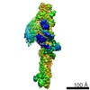 2860MC 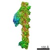 2854C 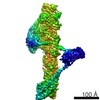 2855C 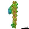 2856C 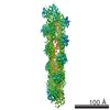 2857C 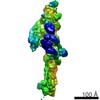 2861C 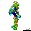 2862C  5adxC 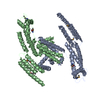 5afrC M: map data used to model this data C: citing same article ( |
|---|---|
| Similar structure data |
-Links
 PDBj
PDBj


- Assembly
| Deposited unit | 
|
|---|---|
| 1 |
|
-Components
-Protein , 16 types, 27 molecules 123456ABCDEFGIHJKLMNOPQRUVb
| #1: Protein | Mass: 30740.773 Da / Num. of mol.: 1 / Source method: isolated from a natural source / Source: (natural) SUS SCROFA (pig) | ||||||||||||||||||||||||||
|---|---|---|---|---|---|---|---|---|---|---|---|---|---|---|---|---|---|---|---|---|---|---|---|---|---|---|---|
| #2: Protein | Mass: 30570.562 Da / Num. of mol.: 1 / Source method: isolated from a natural source / Source: (natural) SUS SCROFA (pig) | ||||||||||||||||||||||||||
| #3: Protein | Mass: 38900.738 Da / Num. of mol.: 2 / Source method: isolated from a natural source / Source: (natural) SUS SCROFA (pig) / References: UniProt: A0A0J9X2A1*PLUS#4: Protein | Mass: 23421.752 Da / Num. of mol.: 2 / Source method: isolated from a natural source / Source: (natural) SUS SCROFA (pig)#5: Protein | Mass: 41959.930 Da / Num. of mol.: 8 / Source method: isolated from a natural source / Source: (natural) SUS SCROFA (pig) / Organ: BRAIN / References: UniProt: F2Z5G5#6: Protein | | / BETA-ACTIN / ACTIN / CYTOPLASMIC 1 / N-TERMINALLY PROCESSEDMass: 41193.043 Da / Num. of mol.: 1 / Source method: isolated from a natural source / Source: (natural) SUS SCROFA (pig) / Organ: BRAIN / References: UniProt: Q6QAQ1#7: Protein | | Mass: 42141.059 Da / Num. of mol.: 1 / Source method: isolated from a natural source / Source: (natural) SUS SCROFA (pig) / Organ: BRAIN / References: UniProt: I3LHK5#8: Protein | | Mass: 31777.492 Da / Num. of mol.: 1 / Source method: isolated from a natural source / Source: (natural) SUS SCROFA (pig) / Organ: BRAIN / References: UniProt: A0PFK5#9: Protein | | Mass: 30509.490 Da / Num. of mol.: 1 / Source method: isolated from a natural source / Source: (natural) SUS SCROFA (pig) / Organ: BRAIN / References: UniProt: D2JYW4#10: Protein | | Mass: 49974.707 Da / Num. of mol.: 1 / Source method: isolated from a natural source / Source: (natural) SUS SCROFA (pig) / Organ: BRAIN#11: Protein | | Mass: 52442.777 Da / Num. of mol.: 1 / Source method: isolated from a natural source / Source: (natural) SUS SCROFA (pig) / Organ: BRAIN#12: Protein | Mass: 5549.833 Da / Num. of mol.: 2 / Source method: isolated from a natural source / Source: (natural) SUS SCROFA (pig) / Organ: BRAIN#13: Protein | Mass: 7422.140 Da / Num. of mol.: 2 / Source method: isolated from a natural source / Source: (natural) SUS SCROFA (pig) / Organ: BRAIN#14: Protein | | Mass: 18343.203 Da / Num. of mol.: 1 / Source method: isolated from a natural source / Source: (natural) SUS SCROFA (pig) / Organ: BRAIN / References: UniProt: D0G6S1*PLUS#15: Protein | | Mass: 18178.328 Da / Num. of mol.: 1 / Source method: isolated from a natural source / Source: (natural) SUS SCROFA (pig) / Organ: BRAIN / References: UniProt: A0A0J9X291*PLUS#19: Protein | | Mass: 7931.814 Da / Num. of mol.: 1 / Source method: isolated from a natural source / Source: (natural) SUS SCROFA (pig) / Organ: BRAIN / References: UniProt: A0A0J9X293*PLUS |
-F-ACTIN-CAPPING PROTEIN SUBUNIT ... , 3 types, 3 molecules YZz
| #16: Protein | Mass: 20698.426 Da / Num. of mol.: 1 / Source method: isolated from a natural source / Source: (natural) SUS SCROFA (pig) / Organ: BRAIN |
|---|---|
| #17: Protein | Mass: 4443.468 Da / Num. of mol.: 1 / Source method: isolated from a natural source / Source: (natural) SUS SCROFA (pig) / Organ: BRAIN |
| #22: Protein | Mass: 4528.573 Da / Num. of mol.: 1 / Source method: isolated from a natural source / Source: (natural) SUS SCROFA (pig) / Organ: BRAIN |
-Protein/peptide , 3 types, 3 molecules acd
| #18: Protein/peptide | Mass: 5400.015 Da / Num. of mol.: 1 / Source method: isolated from a natural source / Source: (natural) SUS SCROFA (pig) / Organ: BRAIN / References: UniProt: A0A0J9X292*PLUS |
|---|---|
| #20: Protein/peptide | Mass: 3019.344 Da / Num. of mol.: 1 / Source method: isolated from a natural source / Source: (natural) SUS SCROFA (pig) / Organ: BRAIN / References: UniProt: A0A0J9X299*PLUS |
| #21: Protein/peptide | Mass: 2237.488 Da / Num. of mol.: 1 / Source method: isolated from a natural source / Source: (natural) SUS SCROFA (pig) / Organ: BRAIN / References: UniProt: A0A0J9X295*PLUS |
-Non-polymers , 2 types, 10 molecules 

| #23: Chemical | ChemComp-ADP / Adenosine diphosphate Mass: 427.201 Da / Num. of mol.: 9 / Source method: obtained synthetically / Formula: C10H15N5O10P2 / Comment: ADP, energy-carrying molecule*YM Mass: 427.201 Da / Num. of mol.: 9 / Source method: obtained synthetically / Formula: C10H15N5O10P2 / Comment: ADP, energy-carrying molecule*YM#24: Chemical | ChemComp-ATP / | Adenosine triphosphate Mass: 507.181 Da / Num. of mol.: 1 / Source method: obtained synthetically / Formula: C10H16N5O13P3 / Comment: ATP, energy-carrying molecule*YM Mass: 507.181 Da / Num. of mol.: 1 / Source method: obtained synthetically / Formula: C10H16N5O13P3 / Comment: ATP, energy-carrying molecule*YM |
|---|
-Experimental details
-Experiment
| Experiment | Method: ELECTRON MICROSCOPY |
|---|---|
| EM experiment | Aggregation state: PARTICLE / 3D reconstruction method: single particle reconstruction |
- Sample preparation
| Component | Name: CRYO-EM STRUCTURE OF DYNEIN TAIL-DYNACTIN- BICD2N COMPLEX Type: COMPLEX Details: THE PARTICLES WERE SELECTED USING AN AUTOMATIC SELECTION PROGRAM. |
|---|---|
| Buffer solution | Name: 150MM KCL, 25MM HEPES-KOH, 1MM MGCL2, 0.1MM MGATP, 5MM DTT pH: 7.4 Details: 150MM KCL, 25MM HEPES-KOH, 1MM MGCL2, 0.1MM MGATP, 5MM DTT |
| Specimen | Conc.: 0.08 mg/ml / Embedding applied: NO / Shadowing applied: NO / Staining applied: NO / Vitrification applied: YES |
| Specimen support | Details: HOLEY CARBON |
| Vitrification | Instrument: FEI VITROBOT MARK III / Cryogen name: ETHANE / Details: LIQUID ETHANE |
- Electron microscopy imaging
| Experimental equipment |  Model: Titan Krios / Image courtesy: FEI Company |
|---|---|
| Microscopy | Model: FEI TITAN KRIOS / Date: Sep 12, 2014 |
| Electron gun | Electron source: FIELD EMISSION GUN / Accelerating voltage: 300 kV / Illumination mode: FLOOD BEAM |
| Electron lens | Mode: BRIGHT FIELDBright-field microscopy / Nominal magnification: 47000 X / Calibrated magnification: 82353 X / Nominal defocus max: 8000 nm / Nominal defocus min: 3000 nm / Cs: 2.7 mm |
| Specimen holder | Temperature: 100 K |
| Image recording | Electron dose: 2 e/Å2 / Film or detector model: FEI FALCON II (4k x 4k) |
| Image scans | Num. digital images: 14423 |
| Radiation wavelength | Relative weight: 1 |
- Processing
| Symmetry | Point symmetry: C1 (asymmetric) | ||||||||||||
|---|---|---|---|---|---|---|---|---|---|---|---|---|---|
| 3D reconstruction | Resolution: 8.2 Å / Resolution method: FSC 0.143 CUT-OFF / Num. of particles: 85744 / Refinement type: HALF-MAPS REFINED INDEPENDENTLY / Symmetry type: POINT | ||||||||||||
| Refinement | Highest resolution: 3.5 Å | ||||||||||||
| Refinement step | Cycle: LAST / Highest resolution: 3.5 Å
|
