 Movie
Movie Controller

[English] 日本語
 Yorodumi
- PDB-5a42: Cryo-EM single particle 3D reconstruction of the native conformat... -
+ Open data

- Basic information
| Entry | Database: PDB / ID: 5a42 | ||||||
|---|---|---|---|---|---|---|---|
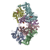
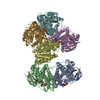
| Title | Cryo-EM single particle 3D reconstruction of the native conformation of E. coli alpha-2-macroglobulin (ECAM) | ||||||
 Components Components | UNCHARACTERIZED LIPOPROTEIN YFHM | ||||||
 Keywords Keywords | HYDROLASE INHIBITOR /  PEPTIDASE INHIBITOR PEPTIDASE INHIBITOR | ||||||
| Function / homology |  Function and homology informationendopeptidase inhibitor activity / protein homodimerization activity / extracellular space / plasma membrane Function and homology informationendopeptidase inhibitor activity / protein homodimerization activity / extracellular space / plasma membraneSimilarity search - Function | ||||||
| Biological species |  ESCHERICHIA COLI K-12 (bacteria) ESCHERICHIA COLI K-12 (bacteria) | ||||||
| Method | ELECTRON MICROSCOPY / single particle reconstruction / cryo EM / Resolution: 16 Å | ||||||
 Authors Authors | Garcia-Ferrer, I. / Arede, P. / Gomez-Blanco, J. / Luque, D. / Duquerroy, S. / Caston, J.R. / Goulas, T. / Gomis-Ruth, F.X. | ||||||
 Citation Citation | Journal: Proc Natl Acad Sci U S A / Year: 2015 Title: Structural and functional insights into Escherichia coli α2-macroglobulin endopeptidase snap-trap inhibition. Authors: Irene Garcia-Ferrer / Pedro Arêde / Josué Gómez-Blanco / Daniel Luque / Stephane Duquerroy / José R Castón / Theodoros Goulas / F Xavier Gomis-Rüth /   Abstract: The survival of commensal bacteria requires them to evade host peptidases. Gram-negative bacteria from the human gut microbiome encode a relative of the human endopeptidase inhibitor, α2- ...The survival of commensal bacteria requires them to evade host peptidases. Gram-negative bacteria from the human gut microbiome encode a relative of the human endopeptidase inhibitor, α2-macroglobulin (α2M). Escherichia coli α2M (ECAM) is a ∼ 180-kDa multidomain membrane-anchored pan-peptidase inhibitor, which is cleaved by host endopeptidases in an accessible bait region. Structural studies by electron microscopy and crystallography reveal that this cleavage causes major structural rearrangement of more than half the 13-domain structure from a native to a compact induced form. It also exposes a reactive thioester bond, which covalently traps the peptidase. Subsequently, peptidase-laden ECAM is shed from the membrane and may dimerize. Trapped peptidases are still active except against very large substrates, so inhibition potentially prevents damage of large cell envelope components, but not host digestion. Mechanistically, these results document a novel monomeric "snap trap." | ||||||
| History |
|
- Structure visualization
| Movie |
Movie viewer |
|---|---|
| Structure viewer | Molecule: MolmilJmol/JSmol |
- Downloads & links
-Download
| PDBx/mmCIF format | 5a42.cif.gz | 275.8 KB | Display | PDBx/mmCIF format |
|---|---|---|---|---|
| PDB format | pdb5a42.ent.gz | 217.8 KB | Display | PDB format |
| PDBx/mmJSON format | 5a42.json.gz | Tree view | PDBx/mmJSON format | |
| Others |  Other downloads Other downloads |
-Validation report
| Arichive directory | https://data.pdbj.org/pub/pdb/validation_reports/a4/5a42ftp://data.pdbj.org/pub/pdb/validation_reports/a4/5a42 | HTTPS FTP |
|---|
-Related structure data
| Related structure data | 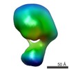 3016MC 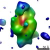 3017C 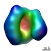 3018C 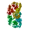 4ziqC 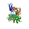 4ziuC 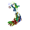 4zjgC 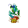 4zjhC M: map data used to model this data C: citing same article ( |
|---|---|
| Similar structure data |


-Links
 PDBj
PDBj


- Assembly
| Deposited unit | 
|
|---|---|
| 1 |
|
-Components
| #1: Protein | Mass: 177690.297 Da / Num. of mol.: 1 / Fragment: RESIDUES 40-1653 Source method: isolated from a genetically manipulated source Details: THE EXPRESSED AND PURIFIED SAMPLE CONTAINED THE SEQUENCE GPM-A40 TO P1653. Source: (gene. exp.) ESCHERICHIA COLI K-12 (bacteria) / Plasmid: PCRI8B / Production host: ESCHERICHIA COLI (E. coli) / Strain (production host): BL21(DE3) / References: UniProt: P76578 |
|---|---|
| Sequence details | THE PURIFIED SAMPLE CONTAINED FROM A40 TO P1653 |
-Experimental details
-Experiment
| Experiment | Method: ELECTRON MICROSCOPY |
|---|---|
| EM experiment | Aggregation state: PARTICLE / 3D reconstruction method: single particle reconstruction |
- Sample preparation
| Component | Name: NATIVE CONFORMATION OF THE E. COLI ALPHA-2- MACROGLOBULIN (ECAM). Type: COMPLEX |
|---|---|
| Buffer solution | Name: 10MM TRIS-HCL, 75MM NACL / pH: 7.4 / Details: 10MM TRIS-HCL, 75MM NACL |
| Specimen | Conc.: 0.01 mg/ml / Embedding applied: NO / Shadowing applied: NO / Staining applied: NO / Vitrification applied: YES |
| Specimen support | Details: CARBON |
| Vitrification | Instrument: LEICA EM CPC / Cryogen name: ETHANE Details: VITRIFICATION 1 -- CRYOGEN- ETHANE, INSTRUMENT- LEICA EM CPC, METHOD- BLOTTED FOR 1MIN BEFORE PLUNGING WITH 5UL PURIFIED SAMPLE., |
- Electron microscopy imaging
| Microscopy | Model: FEI TECNAI 20 / Date: Jun 10, 2014 |
|---|---|
| Electron gun | Electron source: FIELD EMISSION GUN / Accelerating voltage: 200 kV / Illumination mode: FLOOD BEAM |
| Electron lens | Mode: BRIGHT FIELDBright-field microscopy / Calibrated magnification: 84269 X / Nominal defocus max: 3500 nm / Nominal defocus min: 1500 nm |
| Image recording | Film or detector model: FEI EAGLE (4k x 4k) |
| Image scans | Num. digital images: 130 |
| Radiation wavelength | Relative weight: 1 |
- Processing
| EM software |
| ||||||||||||||||
|---|---|---|---|---|---|---|---|---|---|---|---|---|---|---|---|---|---|
| CTF correction | Details: EACH IMAGE | ||||||||||||||||
| Symmetry | Point symmetry: C1 (asymmetric) | ||||||||||||||||
| 3D reconstruction | Method: PROJECTION MATCHING / Resolution: 16 Å / Num. of particles: 46842 / Actual pixel size: 4.5 Å Details: THE FITTED MODEL IS AN HOMOLOGY MODEL BASED ON A ECAM HOMOLOG FROM SALMONELLA ENTERICA (PDB CODE 4U48) AND THE CRYSTAL STRUCTURES ECAM DOMAINS (PDB CODES 4ZIU,4ZJH,4ZJG,4ZIQ). SUBMISSION ...Details: THE FITTED MODEL IS AN HOMOLOGY MODEL BASED ON A ECAM HOMOLOG FROM SALMONELLA ENTERICA (PDB CODE 4U48) AND THE CRYSTAL STRUCTURES ECAM DOMAINS (PDB CODES 4ZIU,4ZJH,4ZJG,4ZIQ). SUBMISSION BASED ON EXPERIMENTAL DATA FROM EMDB EMD-3016. (DEPOSITION ID: 13326). Symmetry type: POINT | ||||||||||||||||
| Atomic model building | Protocol: RIGID BODY FIT / Space: REAL / Target criteria: Cross-correlation coefficient / Details: METHOD--RIGID BODY | ||||||||||||||||
| Refinement | Highest resolution: 16 Å | ||||||||||||||||
| Refinement step | Cycle: LAST / Highest resolution: 16 Å
|
