 Movie
Movie Controller

[English] 日本語
 Yorodumi
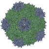
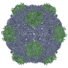
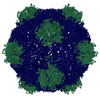
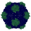
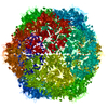
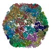
- PDB-5a32: Electron cryo-microscopy of Cowpea Mosaic Virus containing RNA-1 ... -
+ Open data

- Basic information
| Entry | Database: PDB / ID: 5a32 | ||||||
|---|---|---|---|---|---|---|---|
| Title | Electron cryo-microscopy of Cowpea Mosaic Virus containing RNA-1 (CPMVb) | ||||||
 Components Components | (RNA2 POLYPROTEIN) x 2 | ||||||
 Keywords Keywords |  VIRUS / CPMV / COMOVIRIDAE / PICORNAVIRALES. VIRUS / CPMV / COMOVIRIDAE / PICORNAVIRALES. | ||||||
| Function / homology |  Function and homology information Function and homology informationtransport of virus in host, cell to cell / host cell plasmodesma / T=3 icosahedral viral capsid / host cell nucleus / structural molecule activity / GTP binding / DNA binding / RNA bindingSimilarity search - Function | ||||||
| Biological species |  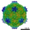 COWPEA MOSAIC VIRUS COWPEA MOSAIC VIRUS | ||||||
| Method | ELECTRON MICROSCOPY / single particle reconstruction / cryo EM / Resolution: 3.44 Å | ||||||
 Authors Authors | Hesketh, E.L. / Meshcheriakova, Y. / Dent, K.C. / Saxena, P. / Thompson, R. / Cockburn, J.J. / Lomonossoff, G.P. / Ranson, N.A. | ||||||
 Citation Citation | Journal: Nat Commun / Year: 2015 Title: Mechanisms of assembly and genome packaging in an RNA virus revealed by high-resolution cryo-EM. Authors: Emma L Hesketh / Yulia Meshcheriakova / Kyle C Dent / Pooja Saxena / Rebecca F Thompson / Joseph J Cockburn / George P Lomonossoff / Neil A Ranson /  Abstract: Cowpea mosaic virus is a plant-infecting member of the Picornavirales and is of major interest in the development of biotechnology applications. Despite the availability of >100 crystal structures of ...Cowpea mosaic virus is a plant-infecting member of the Picornavirales and is of major interest in the development of biotechnology applications. Despite the availability of >100 crystal structures of Picornavirales capsids, relatively little is known about the mechanisms of capsid assembly and genome encapsidation. Here we have determined cryo-electron microscopy reconstructions for the wild-type virus and an empty virus-like particle, to 3.4 Å and 3.0 Å resolution, respectively, and built de novo atomic models of their capsids. These new structures reveal the C-terminal region of the small coat protein subunit, which is essential for virus assembly and which was missing from previously determined crystal structures, as well as residues that bind to the viral genome. These observations allow us to develop a new model for genome encapsidation and capsid assembly. | ||||||
| History |
|
- Structure visualization
| Movie |
Movie viewer |
|---|---|
| Structure viewer | Molecule: MolmilJmol/JSmol |
- Downloads & links
-Download
| PDBx/mmCIF format | 5a32.cif.gz | 117.4 KB | Display | PDBx/mmCIF format |
|---|---|---|---|---|
| PDB format | pdb5a32.ent.gz | 95.3 KB | Display | PDB format |
| PDBx/mmJSON format | 5a32.json.gz | Tree view | PDBx/mmJSON format | |
| Others |  Other downloads Other downloads |
-Validation report
| Arichive directory | https://data.pdbj.org/pub/pdb/validation_reports/a3/5a32ftp://data.pdbj.org/pub/pdb/validation_reports/a3/5a32 | HTTPS FTP |
|---|
-Related structure data
| Related structure data | 3013MC  3014C 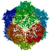 5a33C M: map data used to model this data C: citing same article ( |
|---|---|
| Similar structure data |
-Links
 PDBj
PDBj
- Assembly
| Deposited unit | 
|
|---|---|
| 1 | x 60
|
| 2 |
|
| 3 | x 5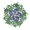
|
| 4 | x 6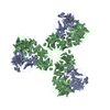
|
| 5 | 
|
| Symmetry | Point symmetry: (Schoenflies symbol: I (icosahedral)) |
-Components
| #1: Protein | Mass: 20961.564 Da / Num. of mol.: 1 / Fragment: LARGE COAT PROTEIN / Source method: isolated from a natural source / Details: WILD-TYPE VIRUS CONTAINING RNA-1 / Source: (natural) COWPEA MOSAIC VIRUS / References: UniProt: P03599 |
|---|---|
| #2: Protein | Mass: 40858.434 Da / Num. of mol.: 1 / Fragment: SMALL COAT PROTEIN N-TERMINUS PART / Source method: isolated from a natural source / Details: WILD-TYPE VIRUS CONTAINING RNA-1 / Source: (natural) COWPEA MOSAIC VIRUS / References: UniProt: P03599 |
-Experimental details
-Experiment
| Experiment | Method: ELECTRON MICROSCOPY |
|---|---|
| EM experiment | Aggregation state: PARTICLE / 3D reconstruction method: single particle reconstruction |
- Sample preparation
| Component | Name: COWPEA MOSAIC VIRUS / Type: VIRUS |
|---|---|
| Specimen | Conc.: 5.8 mg/ml / Embedding applied: NO / Shadowing applied: NO / Staining applied: NO / Vitrification applied: YES |
| Specimen support | Details: HOLEY CARBON |
| Vitrification | Instrument: FEI VITROBOT MARK IV / Cryogen name: ETHANE |
- Electron microscopy imaging
| Experimental equipment |  Model: Titan Krios / Image courtesy: FEI Company |
|---|---|
| Microscopy | Model: FEI TITAN KRIOS / Date: Nov 1, 2014 |
| Electron gun | Electron source: FIELD EMISSION GUN / Accelerating voltage: 300 kV / Illumination mode: FLOOD BEAM |
| Electron lens | Mode: BRIGHT FIELDBright-field microscopy / Nominal magnification: 134615 X / Calibrated magnification: 134615 X / Nominal defocus max: 8000 nm / Nominal defocus min: 500 nm / Cs: 2.7 mm |
| Image recording | Electron dose: 45 e/Å2 / Film or detector model: FEI FALCON II (4k x 4k) |
| Image scans | Num. digital images: 1754 |
- Processing
| EM software | Name: RELION / Version: 1.3 / Category: 3D reconstruction | ||||||||||||
|---|---|---|---|---|---|---|---|---|---|---|---|---|---|
| CTF correction | Details: CTFFIND3 PER MICROGRAPH | ||||||||||||
| Symmetry | Point symmetry: I (icosahedral) | ||||||||||||
| 3D reconstruction | Resolution: 3.44 Å / Num. of particles: 4331 / Nominal pixel size: 1.04 Å / Actual pixel size: 1.04 Å Details: SUBMISSION BASED ON EXPERIMENTAL DATA FROM EMDB EMD-3013. (DEPOSITION ID: 13382). Symmetry type: POINT | ||||||||||||
| Atomic model building | Protocol: FLEXIBLE FIT / Space: REAL / Target criteria: R-factor / Details: REFINEMENT PROTOCOL--EM | ||||||||||||
| Refinement | Highest resolution: 3.44 Å | ||||||||||||
| Refinement step | Cycle: LAST / Highest resolution: 3.44 Å
|
