 Movie
Movie Controller


+ Open data

- Basic information
| Entry | Database: PDB / ID: 4v7q | ||||||||||||
|---|---|---|---|---|---|---|---|---|---|---|---|---|---|
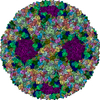
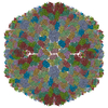
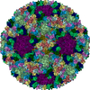
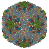
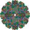
| Title | Atomic model of an infectious rotavirus particle | ||||||||||||
 Components Components |
| ||||||||||||
 Keywords Keywords |  VIRUS / Rotavirus / Triple Layered Particle / Near Atomic Resolution / VP2 / VP6 / VP4 / VP7 / Double layered particle / de novo / Infectious / DLP / ICOSAHEDRAL VIRUS VIRUS / Rotavirus / Triple Layered Particle / Near Atomic Resolution / VP2 / VP6 / VP4 / VP7 / Double layered particle / de novo / Infectious / DLP / ICOSAHEDRAL VIRUS | ||||||||||||
| Function / homology |  Function and homology information Function and homology informationviral intermediate capsid / host cell endoplasmic reticulum lumen / T=13 icosahedral viral capsid / T=2 icosahedral viral capsid / host cell rough endoplasmic reticulum / viral inner capsid / host cytoskeleton / viral outer capsid / permeabilization of host organelle membrane involved in viral entry into host cell / symbiont entry into host cell via permeabilization of inner membrane ...viral intermediate capsid / host cell endoplasmic reticulum lumen / T=13 icosahedral viral capsid / T=2 icosahedral viral capsid / host cell rough endoplasmic reticulum / viral inner capsid / host cytoskeleton / viral outer capsid / permeabilization of host organelle membrane involved in viral entry into host cell / symbiont entry into host cell via permeabilization of inner membrane / host cell endoplasmic reticulum-Golgi intermediate compartment / viral nucleocapsid / receptor-mediated virion attachment to host cell / host cell surface receptor binding / fusion of virus membrane with host plasma membrane / viral envelope / structural molecule activity / virion attachment to host cell / host cell plasma membrane / RNA binding / membrane / metal ion bindingSimilarity search - Function | ||||||||||||
| Biological species |  Simian rotavirus ARhesus rotavirus Simian rotavirus ARhesus rotavirus | ||||||||||||
| Method | ELECTRON MICROSCOPY / single particle reconstruction / cryo EM / Resolution: 3.8 Å | ||||||||||||
 Authors Authors | Settembre, E.C. / Chen, J.Z. / Dormitzer, P.R. / Grigorieff, N. / Harrison, S.C. | ||||||||||||
 Citation Citation | Journal: EMBO J / Year: 2011 Title: Atomic model of an infectious rotavirus particle. Authors: Ethan C Settembre / James Z Chen / Philip R Dormitzer / Nikolaus Grigorieff / Stephen C Harrison /  Abstract: Non-enveloped viruses of different types have evolved distinct mechanisms for penetrating a cellular membrane during infection. Rotavirus penetration appears to occur by a process resembling ...Non-enveloped viruses of different types have evolved distinct mechanisms for penetrating a cellular membrane during infection. Rotavirus penetration appears to occur by a process resembling enveloped-virus fusion: membrane distortion linked to conformational changes in a viral protein. Evidence for such a mechanism comes from crystallographic analyses of fragments of VP4, the rotavirus-penetration protein, and infectivity analyses of structure-based VP4 mutants. We describe here the structure of an infectious rotavirus particle determined by electron cryomicroscopy (cryoEM) and single-particle analysis at about 4.3 Å resolution. The cryoEM image reconstruction permits a nearly complete trace of the VP4 polypeptide chain, including the positions of most side chains. It shows how the two subfragments of VP4 (VP8(*) and VP5(*)) retain their association after proteolytic cleavage, reveals multiple structural roles for the β-barrel domain of VP5(*), and specifies interactions of VP4 with other capsid proteins. The virion model allows us to integrate structural and functional information into a coherent mechanism for rotavirus entry. | ||||||||||||
| History |
|
- Structure visualization
| Movie |
Movie viewer |
|---|---|
| Structure viewer | Molecule: MolmilJmol/JSmol |
- Downloads & links
-Download
| PDBx/mmCIF format | 4v7q.cif.gz | 2.1 MB | Display | PDBx/mmCIF format |
|---|---|---|---|---|
| PDB format | pdb4v7q.ent.gz | Display | PDB format | |
| PDBx/mmJSON format | 4v7q.json.gz | Tree view | PDBx/mmJSON format | |
| Others |  Other downloads Other downloads |
-Validation report
| Arichive directory | https://data.pdbj.org/pub/pdb/validation_reports/v7/4v7qftp://data.pdbj.org/pub/pdb/validation_reports/v7/4v7q | HTTPS FTP |
|---|
-Related structure data
| Related structure data | 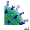 5199MC M: map data used to model this data C: citing same article ( |
|---|---|
| Similar structure data |


-Links
 PDBj
PDBj


- Assembly
| Deposited unit | 
|
|---|---|
| 1 | x 60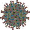
|
| Symmetry | Point symmetry: (Schoenflies symbol: I (icosahedral)) |
-Components
-Protein , 4 types, 31 molecules AAABACADAEAFAGAHAIAJAKALAMANAOBABFBGBHBIBJBKBLBMBNBOBPBQBXBYBZ
| #1: Protein | Mass: 93190.578 Da / Num. of mol.: 2 Source method: isolated from a genetically manipulated source Source: (gene. exp.) Simian rotavirus A / Strain: RRV / Gene: Rotavirus / Production host:  Chlorocebus sabaeus (green monkey) / References: UniProt: B3F2X3 Chlorocebus sabaeus (green monkey) / References: UniProt: B3F2X3#2: Protein | Mass: 44934.766 Da / Num. of mol.: 13 Source method: isolated from a genetically manipulated source Source: (gene. exp.) Rhesus rotavirus / Strain: RRV / Gene: Rotavirus / Production host: Chlorocebus sabaeus (green monkey) / References: UniProt: B2BN53#3: Protein | Mass: 31232.234 Da / Num. of mol.: 13 Source method: isolated from a genetically manipulated source Source: (gene. exp.) Simian rotavirus A / Strain: RRV / Cell (production host): KIDNEY CELLS / Organ (production host): KIDNEY / Production host: Chlorocebus sabaeus (green monkey) / References: UniProt: C3RX25, UniProt: P12476*PLUS#4: Protein | Mass: 86655.586 Da / Num. of mol.: 3 Source method: isolated from a genetically manipulated source Source: (gene. exp.) Simian rotavirus A / Strain: RRV / Cell (production host): KIDNEY CELLS / Organ (production host): KIDNEY / Production host: Chlorocebus sabaeus (green monkey) / References: UniProt: C3RX20 |
|---|
-Sugars , 2 types, 8 molecules 
| #5: Polysaccharide | 2-acetamido-2-deoxy-beta-D-glucopyranose-(1-4)-2-acetamido-2-deoxy-beta-D-glucopyranose #7: Sugar | ChemComp-NAG / | N-Acetylglucosamine Type: D-saccharide, beta linking / Mass: 221.208 Da / Num. of mol.: 1 / Source method: obtained synthetically / Formula: C8H15NO6 Type: D-saccharide, beta linking / Mass: 221.208 Da / Num. of mol.: 1 / Source method: obtained synthetically / Formula: C8H15NO6 |
|---|
-Non-polymers , 1 types, 5 molecules 
| #6: Chemical | ChemComp-ZN /  Mass: 65.409 Da / Num. of mol.: 5 / Source method: obtained synthetically / Formula: Zn Mass: 65.409 Da / Num. of mol.: 5 / Source method: obtained synthetically / Formula: Zn |
|---|
-Experimental details
-Experiment
| Experiment | Method: ELECTRON MICROSCOPY |
|---|---|
| EM experiment | Aggregation state: PARTICLE / 3D reconstruction method: single particle reconstruction |
- Sample preparation
| Component |
| |||||||||||||||
|---|---|---|---|---|---|---|---|---|---|---|---|---|---|---|---|---|
| Details of virus | Host category: VERTEBRATES / Isolate: STRAIN / Type: VIRION | |||||||||||||||
| Natural host | Organism: Macaca mulatta / Strain: Monkey Kidney Cells | |||||||||||||||
| Buffer solution | Name: 20 mM Tris / pH: 7.5 / Details: 20 mM Tris | |||||||||||||||
| Specimen | Conc.: 1 mg/ml / Embedding applied: NO / Shadowing applied: NO / Staining applied: NO / Vitrification applied: YES | |||||||||||||||
| Vitrification | Instrument: HOMEMADE PLUNGER / Cryogen name: ETHANE Details: in normal coldroom environment, ETHANE, manual plunger, front blotting for 3s before plunging, temperature 90 K |
- Electron microscopy imaging
| Experimental equipment |  Model: Tecnai F30 / Image courtesy: FEI Company |
|---|---|
| Microscopy | Model: FEI TECNAI F30 / Date: Mar 1, 2008 Details: Cut-plate film holders to reduce electron back-scattering |
| Electron gun | Electron source: FIELD EMISSION GUN / Accelerating voltage: 300 kV / Illumination mode: FLOOD BEAM |
| Electron lens | Mode: BRIGHT FIELDBright-field microscopy / Nominal magnification: 59000 X / Calibrated magnification: 56772 X / Nominal defocus max: 3000 nm / Nominal defocus min: 1200 nm / Cs: 2 mm |
| Specimen holder | Temperature: 90 K / Tilt angle max: 0 ° / Tilt angle min: 0 ° |
| Image recording | Electron dose: 20 e/Å2 / Film or detector model: KODAK SO-163 FILM |
| Radiation | Protocol: SINGLE WAVELENGTH / Monochromatic (M) / Laue (L): M / Scattering type: x-ray |
| Radiation wavelength | Relative weight: 1 |
- Processing
| CTF correction | Details: individual particle | ||||||||||||
|---|---|---|---|---|---|---|---|---|---|---|---|---|---|
| Symmetry | Point symmetry: I (icosahedral) | ||||||||||||
| 3D reconstruction | Method: projection matching / Resolution: 3.8 Å / Num. of particles: 4187 / Details: icosahedral (I2) averaging / Symmetry type: POINT | ||||||||||||
| Refinement step | Cycle: LAST
|
