 Movie
Movie Controller

[English] 日本語
 Yorodumi
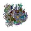
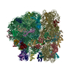
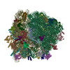
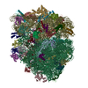
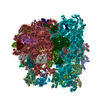
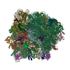
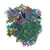
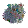
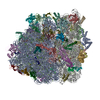
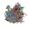
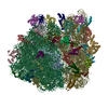
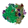
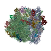
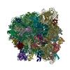
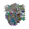
- PDB-4v7c: Structure of the Ribosome with Elongation Factor G Trapped in the... -
+ Open data

- Basic information
| Entry | Database: PDB / ID: 4v7c | |||||||||
|---|---|---|---|---|---|---|---|---|---|---|
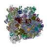
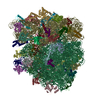
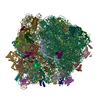
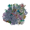
| Title | Structure of the Ribosome with Elongation Factor G Trapped in the Pre-Translocation State (pre-translocation 70S*tRNA structure) | |||||||||
 Components Components |
| |||||||||
 Keywords Keywords | TRANSLATION / EF-G / single particle analysis / pre-translocation translation complex / viomycin | |||||||||
| Function / homology |  Function and homology information Function and homology informationstringent response / ornithine decarboxylase inhibitor activity / transcription antitermination factor activity, RNA binding / misfolded RNA binding / Group I intron splicing / RNA folding / transcriptional attenuation / endoribonuclease inhibitor activity / RNA-binding transcription regulator activity / positive regulation of ribosome biogenesis ...stringent response / ornithine decarboxylase inhibitor activity / transcription antitermination factor activity, RNA binding / misfolded RNA binding / Group I intron splicing / RNA folding / transcriptional attenuation / endoribonuclease inhibitor activity / RNA-binding transcription regulator activity / positive regulation of ribosome biogenesis / negative regulation of cytoplasmic translation / four-way junction DNA binding / translational termination / DnaA-L2 complex / translation repressor activity / negative regulation of translational initiation / negative regulation of DNA-templated DNA replication initiation / regulation of mRNA stability / mRNA regulatory element binding translation repressor activity / ribosome assembly / positive regulation of RNA splicing / assembly of large subunit precursor of preribosome / transcription elongation factor complex / cytosolic ribosome assembly / regulation of DNA-templated transcription elongation / DNA endonuclease activity / ribosomal large subunit assembly / transcription antitermination / response to reactive oxygen species / regulation of cell growth / DNA-templated transcription termination / maintenance of translational fidelity / response to radiation / mRNA 5'-UTR binding / large ribosomal subunit / ribosome biogenesis / ribosome binding / regulation of translation / ribosomal small subunit biogenesis / ribosomal small subunit assembly / small ribosomal subunit / small ribosomal subunit rRNA binding / transferase activity / 5S rRNA binding / large ribosomal subunit rRNA binding / cytosolic small ribosomal subunit / cytosolic large ribosomal subunit / cytoplasmic translation / tRNA binding / molecular adaptor activity / rRNA binding / negative regulation of translation / ribosome / structural constituent of ribosome / translation / response to antibiotic / negative regulation of DNA-templated transcription / mRNA binding / DNA binding / RNA binding / zinc ion binding / membrane / cytoplasm / cytosol Similarity search - Function | |||||||||
| Biological species |  Streptomyces (bacteria) Streptomyces (bacteria) | |||||||||
| Method | ELECTRON MICROSCOPY / single particle reconstruction / cryo EM / Resolution: 7.6 Å | |||||||||
 Authors Authors | Brilot, A.F. / Korostelev, A.A. / Ermolenko, D.N. / Grigorieff, N. | |||||||||
 Citation Citation | Journal: Proc Natl Acad Sci U S A / Year: 2013 Title: Structure of the ribosome with elongation factor G trapped in the pretranslocation state. Authors: Axel F Brilot / Andrei A Korostelev / Dmitri N Ermolenko / Nikolaus Grigorieff /  Abstract: During protein synthesis, tRNAs and their associated mRNA codons move sequentially on the ribosome from the A (aminoacyl) site to the P (peptidyl) site to the E (exit) site in a process catalyzed by ...During protein synthesis, tRNAs and their associated mRNA codons move sequentially on the ribosome from the A (aminoacyl) site to the P (peptidyl) site to the E (exit) site in a process catalyzed by a universally conserved ribosome-dependent GTPase [elongation factor G (EF-G) in prokaryotes and elongation factor 2 (EF-2) in eukaryotes]. Although the high-resolution structure of EF-G bound to the posttranslocation ribosome has been determined, the pretranslocation conformation of the ribosome bound with EF-G and A-site tRNA has evaded visualization owing to the transient nature of this state. Here we use electron cryomicroscopy to determine the structure of the 70S ribosome with EF-G, which is trapped in the pretranslocation state using antibiotic viomycin. Comparison with the posttranslocation ribosome shows that the small subunit of the pretranslocation ribosome is rotated by ∼12° relative to the large subunit. Domain IV of EF-G is positioned in the cleft between the body and head of the small subunit outwardly of the A site and contacts the A-site tRNA. Our findings suggest a model in which domain IV of EF-G promotes the translocation of tRNA from the A to the P site as the small ribosome subunit spontaneously rotates back from the hybrid, rotated state into the nonrotated posttranslocation state. | |||||||||
| History |
|
- Structure visualization
| Movie |
Movie viewer |
|---|---|
| Structure viewer | Molecule: MolmilJmol/JSmol |
- Downloads & links
-Download
| PDBx/mmCIF format | 4v7c.cif.gz | 3.1 MB | Display | PDBx/mmCIF format |
|---|---|---|---|---|
| PDB format | pdb4v7c.ent.gz | Display | PDB format | |
| PDBx/mmJSON format | 4v7c.json.gz | Tree view | PDBx/mmJSON format | |
| Others |  Other downloads Other downloads |
-Validation report
| Summary document | 4v7c_validation.pdf.gz | 1.4 MB | Display | wwPDB validaton report |
|---|---|---|---|---|
| Full document | 4v7c_full_validation.pdf.gz | 1.8 MB | Display | |
| Data in XML | 4v7c_validation.xml.gz | 269.5 KB | Display | |
| Data in CIF | 4v7c_validation.cif.gz | 429.2 KB | Display | |
| Arichive directory | https://data.pdbj.org/pub/pdb/validation_reports/v7/4v7cftp://data.pdbj.org/pub/pdb/validation_reports/v7/4v7c | HTTPS FTP |
-Related structure data
| Related structure data |  5799MC 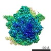 5796C 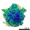 5797C 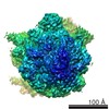 5798C 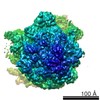 5800C 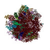 4v7dC M: map data used to model this data C: citing same article ( |
|---|---|
| Similar structure data |

-Links
 PDBj
PDBj


- Assembly
| Deposited unit | 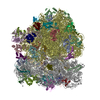
|
|---|---|
| 1 |
|
-Components
-RNA chain , 5 types, 6 molecules AAAVAWAXBABB
| #1: RNA chain | Mass: 499690.031 Da / Num. of mol.: 1 / Source method: isolated from a natural source / Source: (natural) | ||||||
|---|---|---|---|---|---|---|---|
| #22: RNA chain | Mass: 24485.539 Da / Num. of mol.: 2 / Fragment: SEE REMARK 999 / Source method: isolated from a natural source / Source: (natural) #23: RNA chain | | Mass: 5781.499 Da / Num. of mol.: 1 / Fragment: SEE REMARK 999 / Source method: obtained synthetically #25: RNA chain | | Mass: 941306.188 Da / Num. of mol.: 1 / Source method: isolated from a natural source / Source: (natural) #26: RNA chain | | Mass: 38483.926 Da / Num. of mol.: 1 / Source method: isolated from a natural source / Source: (natural) |
-30S ribosomal protein ... , 20 types, 20 molecules ABACADAEAFAGAHAIAJAKALAMANAOAPAQARASATAU
| #2: Protein | Mass: 26650.475 Da / Num. of mol.: 1 / Source method: isolated from a natural source / Source: (natural) |
|---|---|
| #3: Protein | Mass: 25900.117 Da / Num. of mol.: 1 / Source method: isolated from a natural source / Source: (natural) |
| #4: Protein | Mass: 23383.002 Da / Num. of mol.: 1 / Source method: isolated from a natural source / Source: (natural) |
| #5: Protein | Mass: 17498.203 Da / Num. of mol.: 1 / Source method: isolated from a natural source / Source: (natural) |
| #6: Protein | Mass: 15211.058 Da / Num. of mol.: 1 / Source method: isolated from a natural source / Source: (natural) |
| #7: Protein | Mass: 19923.959 Da / Num. of mol.: 1 / Source method: isolated from a natural source / Source: (natural) |
| #8: Protein | Mass: 14015.361 Da / Num. of mol.: 1 / Source method: isolated from a natural source / Source: (natural) |
| #9: Protein | Mass: 14755.074 Da / Num. of mol.: 1 / Source method: isolated from a natural source / Source: (natural) |
| #10: Protein | Mass: 11755.597 Da / Num. of mol.: 1 / Source method: isolated from a natural source / Source: (natural) |
| #11: Protein | Mass: 13739.778 Da / Num. of mol.: 1 / Source method: isolated from a natural source / Source: (natural) |
| #12: Protein | Mass: 13636.961 Da / Num. of mol.: 1 / Source method: isolated from a natural source / Source: (natural) |
| #13: Protein | Mass: 12997.271 Da / Num. of mol.: 1 / Source method: isolated from a natural source / Source: (natural) |
| #14: Protein | Mass: 11475.364 Da / Num. of mol.: 1 / Source method: isolated from a natural source / Source: (natural) |
| #15: Protein | Mass: 10159.621 Da / Num. of mol.: 1 / Source method: isolated from a natural source / Source: (natural) |
| #16: Protein | Mass: 9207.572 Da / Num. of mol.: 1 / Source method: isolated from a natural source / Source: (natural) |
| #17: Protein | Mass: 9593.296 Da / Num. of mol.: 1 / Source method: isolated from a natural source / Source: (natural) |
| #18: Protein | Mass: 8874.276 Da / Num. of mol.: 1 / Source method: isolated from a natural source / Source: (natural) |
| #19: Protein | Mass: 10324.160 Da / Num. of mol.: 1 / Source method: isolated from a natural source / Source: (natural) |
| #20: Protein | Mass: 9577.268 Da / Num. of mol.: 1 / Source method: isolated from a natural source / Source: (natural) |
| #21: Protein | Mass: 8392.844 Da / Num. of mol.: 1 / Source method: isolated from a natural source / Source: (natural) |
+50S ribosomal protein ... , 31 types, 31 molecules BCBDBEBFBGBHBIBJBKBLBMBNBOBPBQBRBSBTBUBVBWBXBYBZB1B2B3B4B5B6B7
-Protein/peptide / Non-polymers , 2 types, 2 molecules AY
| #24: Protein/peptide | 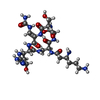  Type: Oligopeptide / Class: Antibiotic / Mass: 703.707 Da / Num. of mol.: 1 / Source method: obtained synthetically / Source: (synth.) Streptomyces (bacteria) / References: NOR: NOR00633, Viomycin Type: Oligopeptide / Class: Antibiotic / Mass: 703.707 Da / Num. of mol.: 1 / Source method: obtained synthetically / Source: (synth.) Streptomyces (bacteria) / References: NOR: NOR00633, Viomycin |
|---|---|
| #58: Chemical | ChemComp-ZN /  Mass: 65.409 Da / Num. of mol.: 1 / Source method: obtained synthetically / Formula: Zn Mass: 65.409 Da / Num. of mol.: 1 / Source method: obtained synthetically / Formula: Zn |
-Details
| Sequence details | THE SEQUENCES OF P/E TRNA (TRNA-MET) AND MRNA (GGCAAGGAGGUAAAAAUGUUUAAACGUAAAUCUACU) USED IN THIS ...THE SEQUENCES OF P/E TRNA (TRNA-MET) AND MRNA (GGCAAGGAGG |
|---|
-Experimental details
-Experiment
| Experiment | Method: ELECTRON MICROSCOPY |
|---|---|
| EM experiment | Aggregation state: PARTICLE / 3D reconstruction method: single particle reconstruction |
- Sample preparation
| Component |
| |||||||||||||||||||||||||
|---|---|---|---|---|---|---|---|---|---|---|---|---|---|---|---|---|---|---|---|---|---|---|---|---|---|---|
| Molecular weight | Value: 3 MDa / Experimental value: NO | |||||||||||||||||||||||||
| Buffer solution | Name: Polymix buffer / pH: 7.6 Details: 10 mM HEPES-KOH, 5 mM MgCl2, 90 mM NH4Cl, 2 mM spermidine, 0.1 mM spermine, 6 mM BME, 0.5 mM viomycin, 0.5 mM GTP, 0.5 mM fusidic acid | |||||||||||||||||||||||||
| Specimen | Conc.: 0.4 mg/ml / Embedding applied: NO / Shadowing applied: NO / Staining applied: NO / Vitrification applied: YES | |||||||||||||||||||||||||
| Specimen support | Details: C-flat 1.2/1.3 holey carbon 400 mesh copper grid, glow discharged with a current of -20 mA for 45 seconds in an EMITECH K100X glow discharge unit | |||||||||||||||||||||||||
| Vitrification | Instrument: FEI VITROBOT MARK II / Cryogen name: ETHANE / Humidity: 95 % Details: Freshly glow-disharged grids were loaded into an FEI Mark II Vitrobot and equilibrated to 95% relative humidity at 22 degrees Celsius. 2 microliters of sample was applied through the side ...Details: Freshly glow-disharged grids were loaded into an FEI Mark II Vitrobot and equilibrated to 95% relative humidity at 22 degrees Celsius. 2 microliters of sample was applied through the side port, blotted for 7 seconds with a positional offset of 2, and plunged into liquid ethane. Method: Freshly glow-disharged grids were loaded into an FEI Mark II Vitrobot and equilibrated to 95% relative humidity at 22 degrees Celsius. 2 microliters of sample was applied through the side ...Method: Freshly glow-disharged grids were loaded into an FEI Mark II Vitrobot and equilibrated to 95% relative humidity at 22 degrees Celsius. 2 microliters of sample was applied through the side port, blotted for 7 seconds with a positional offset of 2, and plunged into liquid ethane. |
- Electron microscopy imaging
| Experimental equipment |  Model: Titan Krios / Image courtesy: FEI Company |
|---|---|
| Microscopy | Model: FEI TITAN KRIOS / Date: Nov 2, 2012 |
| Electron gun | Electron source:  FIELD EMISSION GUN / Accelerating voltage: 300 kV / Illumination mode: FLOOD BEAM FIELD EMISSION GUN / Accelerating voltage: 300 kV / Illumination mode: FLOOD BEAM |
| Electron lens | Mode: BRIGHT FIELD / Nominal magnification: 133333 X / Calibrated magnification: 134615 X / Nominal defocus max: 6950 nm / Nominal defocus min: 1150 nm / Cs: 0.01 mm / Astigmatism: Automatically corrected using FEI software / Camera length: 0 mm |
| Specimen holder | Specimen holder model: FEI TITAN KRIOS AUTOGRID HOLDER |
| Image recording | Electron dose: 30 e/Å2 / Film or detector model: FEI FALCON I (4k x 4k) |
| Image scans | Num. digital images: 13341 |
| Radiation | Protocol: SINGLE WAVELENGTH / Monochromatic (M) / Laue (L): M / Scattering type: x-ray |
| Radiation wavelength | Relative weight: 1 |
- Processing
| EM software |
| ||||||||||||||||||||||||||||||||
|---|---|---|---|---|---|---|---|---|---|---|---|---|---|---|---|---|---|---|---|---|---|---|---|---|---|---|---|---|---|---|---|---|---|
| CTF correction | Details: CTFFIND3, FREALIGN per micrograph | ||||||||||||||||||||||||||||||||
| Symmetry | Point symmetry: C1 (asymmetric) | ||||||||||||||||||||||||||||||||
| 3D reconstruction | Method: Projection Matching / Resolution: 7.6 Å / Resolution method: FSC 0.143 CUT-OFF / Num. of particles: 85115 / Nominal pixel size: 1.05 Å / Actual pixel size: 1.04 Å Details: Refinement included data to 12 Angstrom resolution to limit FSC bias. See primary citation Supplementary Information for details. (Single particle details: Refinement and 3D classification ...Details: Refinement included data to 12 Angstrom resolution to limit FSC bias. See primary citation Supplementary Information for details. (Single particle details: Refinement and 3D classification performed by Frealign) (Single particle--Applied symmetry: C1) Symmetry type: POINT | ||||||||||||||||||||||||||||||||
| Atomic model building | Protocol: RIGID BODY FIT / Space: REAL / Target criteria: Cross-correlation Details: REFINEMENT PROTOCOL--rigid body DETAILS--Chain Y not used in refinement | ||||||||||||||||||||||||||||||||
| Atomic model building | PDB-ID: 4GD1 4gd1 | ||||||||||||||||||||||||||||||||
| Refinement step | Cycle: LAST
|
