 Movie
Movie Controller

[English] 日本語
 Yorodumi
- PDB-4umm: The Cryo-EM structure of the palindromic DNA-bound USP-EcR nuclea... -
+ Open data

- Basic information
| Entry | Database: PDB / ID: 4umm | ||||||
|---|---|---|---|---|---|---|---|
| Title | The Cryo-EM structure of the palindromic DNA-bound USP-EcR nuclear receptor reveals an asymmetric organization with allosteric domain positioning | ||||||
 Components Components |
| ||||||
 Keywords Keywords |  NUCLEAR RECEPTOR / TRANSCRIPTION / ECDYSONE / USP-ECR / DNA RESPONSE ELEMENT / ALLOSTERY / CRYO ELECTRON MICROSCOPY NUCLEAR RECEPTOR / TRANSCRIPTION / ECDYSONE / USP-ECR / DNA RESPONSE ELEMENT / ALLOSTERY / CRYO ELECTRON MICROSCOPY | ||||||
| Function / homology |  Function and homology informationecdysone binding / ecdysone receptor-mediated signaling pathway / nuclear steroid receptor activity / nuclear receptor activity / sequence-specific DNA binding / regulation of DNA-templated transcription / DNA binding / zinc ion binding / nucleus Function and homology informationecdysone binding / ecdysone receptor-mediated signaling pathway / nuclear steroid receptor activity / nuclear receptor activity / sequence-specific DNA binding / regulation of DNA-templated transcription / DNA binding / zinc ion binding / nucleusSimilarity search - Function | ||||||
| Biological species |  HELIOTHIS VIRESCENS (tobacco budworm) HELIOTHIS VIRESCENS (tobacco budworm)SYNTHETIC CONSTRUCT (others) | ||||||
| Method | ELECTRON MICROSCOPY / single particle reconstruction / cryo EM / Resolution: 11.6 Å | ||||||
 Authors Authors | Maletta, M. / Orlov, I. / Moras, D. / Billas, I.M.L. / Klaholz, B.P. | ||||||
 Citation Citation | Journal: Nat Commun / Year: 2014 Title: The palindromic DNA-bound USP/EcR nuclear receptor adopts an asymmetric organization with allosteric domain positioning. Authors: Massimiliano Maletta / Igor Orlov / Pierre Roblin / Yannick Beck / Dino Moras / Isabelle M L Billas / Bruno P Klaholz /  Abstract: Nuclear receptors (NRs) regulate gene expression through DNA- and ligand-binding and thus represent crucial therapeutic targets. The ultraspiracle protein/ecdysone receptor (USP/EcR) complex binds to ...Nuclear receptors (NRs) regulate gene expression through DNA- and ligand-binding and thus represent crucial therapeutic targets. The ultraspiracle protein/ecdysone receptor (USP/EcR) complex binds to half-sites with a one base pair spaced inverted repeat (IR1), a palindromic DNA response element (RE) reminiscent of IRs observed for vertebrate steroid hormone receptors. Here we present the cryo electron microscopy structure of the USP/EcR complex bound to an IR1 RE which provides the first description of a full IR-bound NR complex. The structure reveals that even though the DNA is almost symmetric, the complex adopts a highly asymmetric architecture in which the ligand-binding domains (LBDs) are positioned 5' off-centred. Additional interactions of the USP LBD with the 5'-flanking sequence trigger transcription activity as monitored by transfection assays. The comparison with DR-bound NR complexes suggests that DNA is the major allosteric driver in inversely positioning the LBDs, which serve as the main binding-site for transcriptional regulators. | ||||||
| History |
|
- Structure visualization
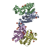
| Movie |
Movie viewer |
|---|---|
| Structure viewer | Molecule: MolmilJmol/JSmol |
- Downloads & links
-Download
| PDBx/mmCIF format | 4umm.cif.gz | 160 KB | Display | PDBx/mmCIF format |
|---|---|---|---|---|
| PDB format | pdb4umm.ent.gz | 125.2 KB | Display | PDB format |
| PDBx/mmJSON format | 4umm.json.gz | Tree view | PDBx/mmJSON format | |
| Others |  Other downloads Other downloads |
-Validation report
| Arichive directory | https://data.pdbj.org/pub/pdb/validation_reports/um/4ummftp://data.pdbj.org/pub/pdb/validation_reports/um/4umm | HTTPS FTP |
|---|
-Related structure data
| Related structure data | 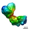 2631MC M: map data used to model this data C: citing same article ( |
|---|---|
| Similar structure data |


-Links
 PDBj
PDBj


- Assembly
| Deposited unit | 
|
|---|---|
| 1 |
|
-Components
-Protein , 4 types, 4 molecules AEFG
| #1: Protein | Mass: 9256.789 Da / Num. of mol.: 1 Source method: isolated from a genetically manipulated source Source: (gene. exp.) HELIOTHIS VIRESCENS (tobacco budworm) / Organ: NUCLEOUS / Production host:  ESCHERICHIA COLI BL21(DE3) (bacteria) ESCHERICHIA COLI BL21(DE3) (bacteria) |
|---|---|
| #4: Protein | / 20-HYDROXY-ECDYSONE RECEPTOR / 20E RECEPTOR / ECRH / ECDYSTEROID RECEPTOR / HVECR / NUCLEAR ...20-HYDROXY-ECDYSONE RECEPTOR / 20E RECEPTOR / ECRH / ECDYSTEROID RECEPTOR / HVECR / NUCLEAR RECEPTOR SUBFAMILY 1 GROUP H MEMBER 1 Mass: 10099.977 Da / Num. of mol.: 1 Source method: isolated from a genetically manipulated source Source: (gene. exp.) HELIOTHIS VIRESCENS (tobacco budworm) / Organ: NUCLEOUS / Production host: ESCHERICHIA COLI BL21(DE3) (bacteria) / References: UniProt: O18473 |
| #5: Protein | Regulation of gene expression / ECR-USP Mass: 29962.717 Da / Num. of mol.: 1 Source method: isolated from a genetically manipulated source Source: (gene. exp.) HELIOTHIS VIRESCENS (tobacco budworm) / Organ: NUCLEOUS / Production host: ESCHERICHIA COLI BL21(DE3) (bacteria) / References: UniProt: Q7SIF6 |
| #6: Protein | / 20-HYDROXY-ECDYSONE RECEPTOR / 20E RECEPTOR / ECRH / ECDYSTEROID RECEPTOR / HVECR / NUCLEAR ...20-HYDROXY-ECDYSONE RECEPTOR / 20E RECEPTOR / ECRH / ECDYSTEROID RECEPTOR / HVECR / NUCLEAR RECEPTOR SUBFAMILY 1 GROUP H MEMBER 1 Mass: 30368.961 Da / Num. of mol.: 1 Source method: isolated from a genetically manipulated source Source: (gene. exp.) HELIOTHIS VIRESCENS (tobacco budworm) / Organ: NUCLEOUS / Production host: ESCHERICHIA COLI BL21(DE3) (bacteria) / References: UniProt: O18473 |
-DNA chain , 2 types, 2 molecules CD
| #2: DNA chain | Mass: 6148.989 Da / Num. of mol.: 1 / Source method: obtained synthetically / Source: (synth.) SYNTHETIC CONSTRUCT (others) |
|---|---|
| #3: DNA chain | Mass: 6117.979 Da / Num. of mol.: 1 / Source method: obtained synthetically / Source: (synth.) SYNTHETIC CONSTRUCT (others) |
-Non-polymers , 3 types, 10 molecules 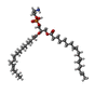

| #7: Chemical | ChemComp-EPH / Phosphatidylethanolamine Mass: 709.933 Da / Num. of mol.: 1 / Source method: obtained synthetically / Formula: C39H68NO8P / Comment: phospholipid*YM Mass: 709.933 Da / Num. of mol.: 1 / Source method: obtained synthetically / Formula: C39H68NO8P / Comment: phospholipid*YM |
|---|---|
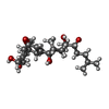
| #8: Chemical | ChemComp-P1A /  Mass: 464.635 Da / Num. of mol.: 1 / Source method: obtained synthetically / Formula: C27H44O6 Mass: 464.635 Da / Num. of mol.: 1 / Source method: obtained synthetically / Formula: C27H44O6 |
| #9: Water | ChemComp-HOH / WaterMass: 18.015 Da / Num. of mol.: 8 / Source method: isolated from a natural source / Formula: H2O |
-Experimental details
-Experiment
| Experiment | Method: ELECTRON MICROSCOPY |
|---|---|
| EM experiment | Aggregation state: PARTICLE / 3D reconstruction method: single particle reconstruction |
- Sample preparation
| Component | Name: BRUNO P. KLAHOLZ / Type: COMPLEX |
|---|---|
| Buffer solution | Name: 10 MM TRIS PH 7.5, 100 MM NACL, 10 MM MGCL2, 10 MM TCEP pH: 7.5 Details: 10 MM TRIS PH 7.5, 100 MM NACL, 10 MM MGCL2, 10 MM TCEP |
| Specimen | Conc.: 0.1 mg/ml / Embedding applied: NO / Shadowing applied: NO / Staining applied: NO / Vitrification applied: YES |
| Specimen support | Details: CARBON |
| Vitrification | Instrument: FEI VITROBOT MARK IV / Cryogen name: ETHANE Details: VITRIFICATION 1 -- CRYOGEN- ETHANE, HUMIDITY- 90, TEMPERATURE- 120, INSTRUMENT- FEI VITROBOT MARK IV, METHOD- 2 SECONDS, FORCE 4, |
- Electron microscopy imaging
| Experimental equipment |  Model: Tecnai F30 / Image courtesy: FEI Company |
|---|---|
| Microscopy | Model: FEI TECNAI F30 / Date: Nov 20, 2009 |
| Electron gun | Electron source: FIELD EMISSION GUN / Accelerating voltage: 100 kV / Illumination mode: FLOOD BEAM |
| Electron lens | Mode: BRIGHT FIELDBright-field microscopy / Nominal magnification: 59000 X / Calibrated magnification: 64244 X / Nominal defocus max: 3000 nm / Nominal defocus min: 1500 nm / Cs: 2 mm |
| Image recording | Electron dose: 20 e/Å2 / Film or detector model: FEI EAGLE (4k x 4k) |
| Image scans | Num. digital images: 600 |
- Processing
| EM software | Name: IMAGIC / Version: 5 / Category: 3D reconstruction | ||||||||||||
|---|---|---|---|---|---|---|---|---|---|---|---|---|---|
| CTF correction | Details: CCD IMAGES 4096X4096 | ||||||||||||
| Symmetry | Point symmetry: C1 (asymmetric) | ||||||||||||
| 3D reconstruction | Method: CROSS-COMMON LINES / Resolution: 11.6 Å / Num. of particles: 50000 Details: SUBMISSION BASED ON EXPERIMENTAL DATA FROM EMDB EMD -2631. (DEPOSITION ID: 12447). Symmetry type: POINT | ||||||||||||
| Atomic model building | Protocol: RIGID BODY FIT / Space: REAL / Details: METHOD--RIGID BODY | ||||||||||||
| Atomic model building | PDB-ID: 1R1K | ||||||||||||
| Refinement | Highest resolution: 11.6 Å | ||||||||||||
| Refinement step | Cycle: LAST / Highest resolution: 11.6 Å
|

