 Movie
Movie Controller


+ Open data

- Basic information
| Entry | Database: PDB / ID: 4uft | ||||||
|---|---|---|---|---|---|---|---|
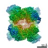
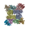
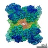
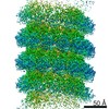
| Title | Structure of the helical Measles virus nucleocapsid | ||||||
 Components Components |
| ||||||
 Keywords Keywords |  RNA BINDING PROTEIN / MEASLES VIRUS NUCLEOCAPSID / TRANSCRIPTION AND REPLICATION TEMPLATE RNA BINDING PROTEIN / MEASLES VIRUS NUCLEOCAPSID / TRANSCRIPTION AND REPLICATION TEMPLATE | ||||||
| Function / homology |  Function and homology information Function and homology informationhelical viral capsid / viral process / viral nucleocapsid / host cell cytoplasm / structural molecule activity / RNA bindingSimilarity search - Function | ||||||
| Biological species |  MEASLES VIRUS STRAIN HALLE MEASLES VIRUS STRAIN HALLE  SPODOPTERA FRUGIPERDA (fall armyworm) SPODOPTERA FRUGIPERDA (fall armyworm) | ||||||
| Method | ELECTRON MICROSCOPY / single particle reconstruction / cryo EM / Resolution: 4.3 Å | ||||||
 Authors Authors | Gutsche, I. / Desfosses, A. / Effantin, G. / Ling, W.L. / Haupt, M. / Ruigrok, R.W.H. / Sachse, C. / Schoehn, G. | ||||||
 Citation Citation | Journal: Science / Year: 2015 Title: Structural virology. Near-atomic cryo-EM structure of the helical measles virus nucleocapsid. Authors: Irina Gutsche / Ambroise Desfosses / Grégory Effantin / Wai Li Ling / Melina Haupt / Rob W H Ruigrok / Carsten Sachse / Guy Schoehn /   Abstract: Measles is a highly contagious human disease. We used cryo-electron microscopy and single particle-based helical image analysis to determine the structure of the helical nucleocapsid formed by the ...Measles is a highly contagious human disease. We used cryo-electron microscopy and single particle-based helical image analysis to determine the structure of the helical nucleocapsid formed by the folded domain of the measles virus nucleoprotein encapsidating an RNA at a resolution of 4.3 angstroms. The resulting pseudoatomic model of the measles virus nucleocapsid offers important insights into the mechanism of the helical polymerization of nucleocapsids of negative-strand RNA viruses, in particular via the exchange subdomains of the nucleoprotein. The structure reveals the mode of the nucleoprotein-RNA interaction and explains why each nucleoprotein of measles virus binds six nucleotides, whereas the respiratory syncytial virus nucleoprotein binds seven. It provides a rational basis for further analysis of measles virus replication and transcription, and reveals potential targets for drug design. | ||||||
| History |
|
- Structure visualization
| Movie |
Movie viewer |
|---|---|
| Structure viewer | Molecule: MolmilJmol/JSmol |
- Downloads & links
-Download
| PDBx/mmCIF format | 4uft.cif.gz | 74.5 KB | Display | PDBx/mmCIF format |
|---|---|---|---|---|
| PDB format | pdb4uft.ent.gz | 57.3 KB | Display | PDB format |
| PDBx/mmJSON format | 4uft.json.gz | Tree view | PDBx/mmJSON format | |
| Others |  Other downloads Other downloads |
-Validation report
| Arichive directory | https://data.pdbj.org/pub/pdb/validation_reports/uf/4uftftp://data.pdbj.org/pub/pdb/validation_reports/uf/4uft | HTTPS FTP |
|---|
-Related structure data
| Related structure data | 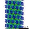 2867MC M: map data used to model this data C: citing same article ( |
|---|---|
| Similar structure data |


-Links
 PDBj
PDBj

- Assembly
| Deposited unit | 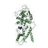
|
|---|---|
| 1 | x 39
|
| 2 |
|
| 3 | 
|
| Symmetry | Helical symmetry: (Circular symmetry: 1 / Dyad axis: no / N subunits divisor: 1 / Num. of operations: 39 / Rise per n subunits: 4.015 Å / Rotation per n subunits: -29.173 °) |
-Components
| #1: Protein | Mass: 43506.621 Da / Num. of mol.: 1 Source method: isolated from a genetically manipulated source Details: NUCLEOPROTEIN WAS PARTIALLY DIGESTED WITH TRYPSIN / Source: (gene. exp.) MEASLES VIRUS STRAIN HALLE / Cell line (production host): Sf21 / Production host: SPODOPTERA FRUGIPERDA (fall armyworm) / References: UniProt: P10050 |
|---|---|
| #2: RNA chain | Mass: 1786.133 Da / Num. of mol.: 1 / Source method: isolated from a natural source / Source: (natural) SPODOPTERA FRUGIPERDA (fall armyworm) |
-Experimental details
-Experiment
| Experiment | Method: ELECTRON MICROSCOPY |
|---|---|
| EM experiment | Aggregation state: PARTICLE / 3D reconstruction method: single particle reconstruction |
- Sample preparation
| Component | Name: RECOMBINANT MEASLES NUCLEOPROTEIN-RNA HELICAL ASSEMBLY (NUCLEOCAPSID) Type: COMPLEX |
|---|---|
| Buffer solution | Name: IN 20 MM TRIHCL PH 7.5, 150 MM NACL / pH: 7.5 / Details: IN 20 MM TRIHCL PH 7.5, 150 MM NACL |
| Specimen | Embedding applied: NO / Shadowing applied: NO / Staining applied: NO / Vitrification applied: YES |
| Specimen support | Details: HOLEY CARBON |
| Vitrification | Instrument: FEI VITROBOT MARK IV / Cryogen name: ETHANE |
- Electron microscopy imaging
| Experimental equipment |  Model: Tecnai F30 / Image courtesy: FEI Company |
|---|---|
| Microscopy | Model: FEI TECNAI F30 / Date: Jul 1, 2013 Details: SPECIAL CARE WAS TAKEN TO PERFORM A COMA-FREE ALIGNMENT OF THE MICROSCOPE |
| Electron gun | Electron source: FIELD EMISSION GUN / Accelerating voltage: 300 kV / Illumination mode: FLOOD BEAM |
| Electron lens | Mode: BRIGHT FIELDBright-field microscopy / Nominal magnification: 59000 X / Nominal defocus max: 3500 nm / Nominal defocus min: 800 nm |
| Image recording | Film or detector model: KODAK SO-163 FILM |
| Radiation wavelength | Relative weight: 1 |
- Processing
| EM software |
| ||||||||||||||||||
|---|---|---|---|---|---|---|---|---|---|---|---|---|---|---|---|---|---|---|---|
| CTF correction | Details: EACH PARTICLE | ||||||||||||||||||
| 3D reconstruction | Method: WEIGHTED BACK-PROJECTION / Resolution: 4.3 Å / Num. of particles: 228165 / Nominal pixel size: 1.186 Å / Actual pixel size: 1.186 Å Details: SUBMISSION BASED ON EXPERIMENTAL DATA FROM EMDB EMD -2867. (DEPOSITION ID: 13046). Symmetry type: HELICAL | ||||||||||||||||||
| Atomic model building | Protocol: FLEXIBLE FIT / Space: REAL / Details: METHOD--FLEXIBLE REFINEMENT PROTOCOL--X-RAY | ||||||||||||||||||
| Atomic model building | PDB-ID: 2WJ8 | ||||||||||||||||||
| Refinement | Highest resolution: 4.3 Å | ||||||||||||||||||
| Refinement step | Cycle: LAST / Highest resolution: 4.3 Å
|

