 Movie
Movie Controller


+ Open data

- Basic information
| Entry | Database: PDB / ID: 4ci0 | ||||||
|---|---|---|---|---|---|---|---|
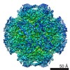
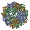
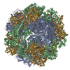
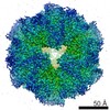
| Title | Electron cryo-microscopy of F420-reducing NiFe hydrogenase Frh | ||||||
 Components Components | (F420-REDUCING HYDROGENASE, SUBUNIT ...) x 3 | ||||||
 Keywords Keywords |  OXIDOREDUCTASE / FLAVOPROTEIN / ELECTRON TRANSFER / FERREDOXIN OXIDOREDUCTASE / FLAVOPROTEIN / ELECTRON TRANSFER / FERREDOXIN | ||||||
| Function / homology |  Function and homology informationcoenzyme F420 hydrogenase / coenzyme F420 hydrogenase activity / ferredoxin hydrogenase activity / iron-sulfur cluster binding / nickel cation binding / flavin adenine dinucleotide binding / 4 iron, 4 sulfur cluster binding Function and homology informationcoenzyme F420 hydrogenase / coenzyme F420 hydrogenase activity / ferredoxin hydrogenase activity / iron-sulfur cluster binding / nickel cation binding / flavin adenine dinucleotide binding / 4 iron, 4 sulfur cluster bindingSimilarity search - Function | ||||||
| Biological species |   METHANOTHERMOBACTER MARBURGENSIS (archaea) METHANOTHERMOBACTER MARBURGENSIS (archaea) | ||||||
| Method | ELECTRON MICROSCOPY / single particle reconstruction / cryo EM / Resolution: 3.36 Å | ||||||
 Authors Authors | Allegretti, M. / Mills, D.J. / McMullan, G. / Kuehlbrandt, W. / Vonck, J. | ||||||
 Citation Citation | Journal: Elife / Year: 2014 Title: Atomic model of the F420-reducing [NiFe] hydrogenase by electron cryo-microscopy using a direct electron detector. Authors: Matteo Allegretti / Deryck J Mills / Greg McMullan / Werner Kühlbrandt / Janet Vonck /  Abstract: The introduction of direct electron detectors with higher detective quantum efficiency and fast read-out marks the beginning of a new era in electron cryo-microscopy. Using the FEI Falcon II direct ...The introduction of direct electron detectors with higher detective quantum efficiency and fast read-out marks the beginning of a new era in electron cryo-microscopy. Using the FEI Falcon II direct electron detector in video mode, we have reconstructed a map at 3.36 Å resolution of the 1.2 MDa F420-reducing hydrogenase (Frh) from methanogenic archaea from only 320,000 asymmetric units. Videos frames were aligned by a combination of image and particle alignment procedures to overcome the effects of beam-induced motion. The reconstructed density map shows all secondary structure as well as clear side chain densities for most residues. The full coordination of all cofactors in the electron transfer chain (a [NiFe] center, four [4Fe4S] clusters and an FAD) is clearly visible along with a well-defined substrate access channel. From the rigidity of the complex we conclude that catalysis is diffusion-limited and does not depend on protein flexibility or conformational changes. DOI: http://dx.doi.org/10.7554/eLife.01963.001. | ||||||
| History |
|
- Structure visualization
| Movie |
Movie viewer |
|---|---|
| Structure viewer | Molecule: MolmilJmol/JSmol |
- Downloads & links
-Download
| PDBx/mmCIF format | 4ci0.cif.gz | 170.3 KB | Display | PDBx/mmCIF format |
|---|---|---|---|---|
| PDB format | pdb4ci0.ent.gz | 136.1 KB | Display | PDB format |
| PDBx/mmJSON format | 4ci0.json.gz | Tree view | PDBx/mmJSON format | |
| Others |  Other downloads Other downloads |
-Validation report
| Arichive directory | https://data.pdbj.org/pub/pdb/validation_reports/ci/4ci0ftp://data.pdbj.org/pub/pdb/validation_reports/ci/4ci0 | HTTPS FTP |
|---|
-Related structure data
| Related structure data |  2513MC M: map data used to model this data C: citing same article ( |
|---|---|
| Similar structure data |

-Links
 PDBj
PDBj


- Assembly
| Deposited unit | 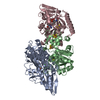
|
|---|---|
| 1 | x 12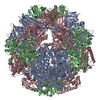
|
| 2 |
|
| 3 | 
|
| Symmetry | Point symmetry: (Schoenflies symbol: T (tetrahedral)) |
-Components
-F420-REDUCING HYDROGENASE, SUBUNIT ... , 3 types, 3 molecules ABC
| #1: Protein | Mass: 42696.773 Da / Num. of mol.: 1 / Fragment: RESIDUES 1-386 / Source method: isolated from a natural source Source: (natural) METHANOTHERMOBACTER MARBURGENSIS (archaea)References: UniProt: D9PYF9, coenzyme F420 hydrogenase |
|---|---|
| #2: Protein | Mass: 30267.762 Da / Num. of mol.: 1 / Source method: isolated from a natural source Source: (natural) METHANOTHERMOBACTER MARBURGENSIS (archaea)References: UniProt: D9PYF7, coenzyme F420 hydrogenase |
| #3: Protein | Mass: 30778.752 Da / Num. of mol.: 1 / Source method: isolated from a natural source Source: (natural) METHANOTHERMOBACTER MARBURGENSIS (archaea)References: UniProt: D9PYF6, coenzyme F420 hydrogenase |
-Non-polymers , 6 types, 9 molecules 


| #4: Chemical | ChemComp-FE / Iron Mass: 55.845 Da / Num. of mol.: 1 / Source method: obtained synthetically / Formula: Fe Mass: 55.845 Da / Num. of mol.: 1 / Source method: obtained synthetically / Formula: Fe | ||||
|---|---|---|---|---|---|
| #5: Chemical | ChemComp-NI / Nickel Mass: 58.693 Da / Num. of mol.: 1 / Source method: obtained synthetically / Formula: Ni Mass: 58.693 Da / Num. of mol.: 1 / Source method: obtained synthetically / Formula: Ni | ||||
| #6: Chemical | ChemComp-FE2 /  Mass: 55.845 Da / Num. of mol.: 1 / Source method: obtained synthetically / Formula: Fe Mass: 55.845 Da / Num. of mol.: 1 / Source method: obtained synthetically / Formula: Fe | ||||
| #7: Chemical | ChemComp-SF4 / Iron–sulfur cluster Mass: 351.640 Da / Num. of mol.: 4 / Source method: obtained synthetically / Formula: Fe4S4 Mass: 351.640 Da / Num. of mol.: 4 / Source method: obtained synthetically / Formula: Fe4S4#8: Chemical | ChemComp-ZN / |  Mass: 65.409 Da / Num. of mol.: 1 / Source method: obtained synthetically / Formula: Zn Mass: 65.409 Da / Num. of mol.: 1 / Source method: obtained synthetically / Formula: Zn#9: Chemical | ChemComp-FAD / | Flavin adenine dinucleotide Mass: 785.550 Da / Num. of mol.: 1 / Source method: obtained synthetically / Formula: C27H33N9O15P2 / Comment: FAD*YM Mass: 785.550 Da / Num. of mol.: 1 / Source method: obtained synthetically / Formula: C27H33N9O15P2 / Comment: FAD*YM |
-Details
| Nonpolymer details | ZINC ION (ZN): A ZINC ION IS SHARED BY TWO FRHG SUBUNITS |
|---|---|
| Sequence details | THE SEQUENCE AFTER HIS386 HAS BEEN REMOVED BY AN ENDOPEPTID |
-Experimental details
-Experiment
| Experiment | Method: ELECTRON MICROSCOPY |
|---|---|
| EM experiment | Aggregation state: PARTICLE / 3D reconstruction method: single particle reconstruction |
- Sample preparation
| Component | Name: F420 REDUCING HYDROGENASE / Type: COMPLEX |
|---|---|
| Buffer solution | Name: 50MM TRIS-HCL, 0.025MM FAD / pH: 7.6 / Details: 50MM TRIS-HCL, 0.025MM FAD |
| Specimen | Conc.: 0.7 mg/ml / Embedding applied: NO / Shadowing applied: NO / Staining applied: NO / Vitrification applied: YES |
| Specimen support | Details: HOLEY CARBON |
| Vitrification | Instrument: FEI VITROBOT MARK I / Cryogen name: ETHANE Details: VITRIFICATION 1 -- CRYOGEN- ETHANE, HUMIDITY- 70, TEMPERATURE- 103, INSTRUMENT- FEI VITROBOT MARK I, METHOD- BLOTTING 2.5 SECONDS BEFORE PLUNGING, |
- Electron microscopy imaging
| Experimental equipment |  Model: Tecnai Polara / Image courtesy: FEI Company |
|---|---|
| Microscopy | Model: FEI POLARA 300 / Date: May 27, 2013 / Details: DATA WAS COLLECTED IN MOVIE MODE |
| Electron gun | Electron source: FIELD EMISSION GUN / Accelerating voltage: 300 kV / Illumination mode: FLOOD BEAM |
| Electron lens | Mode: BRIGHT FIELDBright-field microscopy / Nominal magnification: 78000 X / Calibrated magnification: 106000 X / Nominal defocus max: 2500 nm / Nominal defocus min: 800 nm / Cs: 2.2 mm |
| Specimen holder | Temperature: 79 K |
| Image recording | Electron dose: 22 e/Å2 / Film or detector model: FEI FALCON II (4k x 4k) |
| Image scans | Num. digital images: 235 |
| Radiation wavelength | Relative weight: 1 |
- Processing
| EM software |
| ||||||||||||
|---|---|---|---|---|---|---|---|---|---|---|---|---|---|
| CTF correction | Details: EACH IMAGE | ||||||||||||
| Symmetry | Point symmetry: T (tetrahedral) | ||||||||||||
| 3D reconstruction | Method: MAXIMUM LIKELIHOOD / Resolution: 3.36 Å / Num. of particles: 26000 / Nominal pixel size: 1.32 Å / Actual pixel size: 1.32 Å / Magnification calibration: FIT TO ATOMIC MODEL Details: FRH IS A FRHABG DODECAMER WITH TETRAHEDRAL SYMMETRY. SUBMISSION BASED ON EXPERIMENTAL DATA FROM EMDB EMD-2513. (DEPOSITION ID: 12073). Symmetry type: POINT | ||||||||||||
| Atomic model building | Protocol: FLEXIBLE FIT / Space: REAL / Details: METHOD--FLEXIBLE REFINEMENT PROTOCOL--CRYO-EM | ||||||||||||
| Atomic model building | PDB-ID: 3ZFS | ||||||||||||
| Refinement | Highest resolution: 3.36 Å | ||||||||||||
| Refinement step | Cycle: LAST / Highest resolution: 3.36 Å
|

