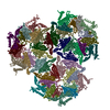
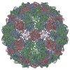
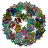
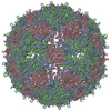
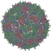
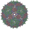
Journal: Structure / Year: 2013 Title: The asymmetric structure of an icosahedral virus bound to its receptor suggests a mechanism for genome release. Authors: Kyle C Dent / Rebecca Thompson / Amy M Barker / Julian A Hiscox / John N Barr / Peter G Stockley / Neil A Ranson / Abstract: Simple, spherical RNA viruses have well-understood, symmetric protein capsids, but little structural information is available for their asymmetric components, such as minor proteins and their ...Simple, spherical RNA viruses have well-understood, symmetric protein capsids, but little structural information is available for their asymmetric components, such as minor proteins and their genomes, which are vital for infection. Here, we report an asymmetric structure of bacteriophage MS2, attached to its receptor, the F-pilus. Cryo-electron tomography and subtomographic averaging of such complexes result in a structure containing clear density for the packaged genome, implying that the conformation of the genome is the same in each virus particle. The data also suggest that the single-copy viral maturation protein breaks the symmetry of the capsid, occupying a position that would be filled by a coat protein dimer in an icosahedral shell. This capsomere can thus fulfill its known biological roles in receptor and genome binding and suggests an exit route for the genome during infection.
History
Deposition
May 23, 2013
Deposition site: PDBE / Processing site: PDBE
Revision 1.0
Jul 17, 2013
Provider: repository / Type: Initial release
Revision 1.1
Sep 11, 2013
Group: Database references / Other
Supersession
Dec 10, 2014
ID: 4BP4, 4BP5, 4BP6 / Details: superseding all the associated split entries
#200 - Aug 2016 Quasisymmetry in Icosahedral Viruses similarity (1)
-
Assembly
Deposited unit
A0: COAT PROTEIN A1: COAT PROTEIN A2: COAT PROTEIN A3: COAT PROTEIN A4: COAT PROTEIN A5: COAT PROTEIN A6: COAT PROTEIN A7: COAT PROTEIN A8: COAT PROTEIN A9: COAT PROTEIN AA: COAT PROTEIN AB: COAT PROTEIN AC: COAT PROTEIN AD: COAT PROTEIN AE: COAT PROTEIN AF: COAT PROTEIN AG: COAT PROTEIN AH: COAT PROTEIN AI: COAT PROTEIN AJ: COAT PROTEIN AK: COAT PROTEIN AL: COAT PROTEIN AM: COAT PROTEIN AN: COAT PROTEIN AO: COAT PROTEIN AP: COAT PROTEIN AQ: COAT PROTEIN AR: COAT PROTEIN AS: COAT PROTEIN AT: COAT PROTEIN AU: COAT PROTEIN AV: COAT PROTEIN AW: COAT PROTEIN AX: COAT PROTEIN AY: COAT PROTEIN AZ: COAT PROTEIN Aa: COAT PROTEIN Ab: COAT PROTEIN Ac: COAT PROTEIN Ad: COAT PROTEIN Ae: COAT PROTEIN Af: COAT PROTEIN Ag: COAT PROTEIN Ah: COAT PROTEIN Ai: COAT PROTEIN Aj: COAT PROTEIN Ak: COAT PROTEIN Al: COAT PROTEIN Am: COAT PROTEIN An: COAT PROTEIN Ao: COAT PROTEIN Ap: COAT PROTEIN Aq: COAT PROTEIN Ar: COAT PROTEIN As: COAT PROTEIN At: COAT PROTEIN Au: COAT PROTEIN Av: COAT PROTEIN Aw: COAT PROTEIN Ax: COAT PROTEIN B0: COAT PROTEIN B1: COAT PROTEIN B2: COAT PROTEIN B3: COAT PROTEIN B4: COAT PROTEIN B5: COAT PROTEIN B6: COAT PROTEIN B7: COAT PROTEIN B8: COAT PROTEIN B9: COAT PROTEIN BA: COAT PROTEIN BB: COAT PROTEIN BC: COAT PROTEIN BD: COAT PROTEIN BE: COAT PROTEIN BF: COAT PROTEIN BG: COAT PROTEIN BH: COAT PROTEIN BI: COAT PROTEIN BJ: COAT PROTEIN BK: COAT PROTEIN BL: COAT PROTEIN BM: COAT PROTEIN BN: COAT PROTEIN BO: COAT PROTEIN BP: COAT PROTEIN BQ: COAT PROTEIN BR: COAT PROTEIN BS: COAT PROTEIN BT: COAT PROTEIN BU: COAT PROTEIN BV: COAT PROTEIN BW: COAT PROTEIN BX: COAT PROTEIN BY: COAT PROTEIN BZ: COAT PROTEIN Ba: COAT PROTEIN Bb: COAT PROTEIN Bc: COAT PROTEIN Bd: COAT PROTEIN Be: COAT PROTEIN Bf: COAT PROTEIN Bg: COAT PROTEIN Bh: COAT PROTEIN Bi: COAT PROTEIN Bj: COAT PROTEIN Bk: COAT PROTEIN Bl: COAT PROTEIN Bm: COAT PROTEIN Bn: COAT PROTEIN Bo: COAT PROTEIN Bp: COAT PROTEIN Bq: COAT PROTEIN Br: COAT PROTEIN Bs: COAT PROTEIN Bt: COAT PROTEIN Bu: COAT PROTEIN Bv: COAT PROTEIN Bw: COAT PROTEIN Bx: COAT PROTEIN C0: COAT PROTEIN C1: COAT PROTEIN C2: COAT PROTEIN C3: COAT PROTEIN C4: COAT PROTEIN C5: COAT PROTEIN C6: COAT PROTEIN C7: COAT PROTEIN C8: COAT PROTEIN C9: COAT PROTEIN CA: COAT PROTEIN CB: COAT PROTEIN CC: COAT PROTEIN CD: COAT PROTEIN CE: COAT PROTEIN CF: COAT PROTEIN CG: COAT PROTEIN CH: COAT PROTEIN CI: COAT PROTEIN CJ: COAT PROTEIN CK: COAT PROTEIN CL: COAT PROTEIN CM: COAT PROTEIN CN: COAT PROTEIN CO: COAT PROTEIN CP: COAT PROTEIN CQ: COAT PROTEIN CR: COAT PROTEIN CS: COAT PROTEIN CT: COAT PROTEIN CU: COAT PROTEIN CV: COAT PROTEIN CW: COAT PROTEIN CX: COAT PROTEIN CY: COAT PROTEIN CZ: COAT PROTEIN Ca: COAT PROTEIN Cb: COAT PROTEIN Cc: COAT PROTEIN Cd: COAT PROTEIN Ce: COAT PROTEIN Cf: COAT PROTEIN Cg: COAT PROTEIN Ch: COAT PROTEIN Ci: COAT PROTEIN Cj: COAT PROTEIN Ck: COAT PROTEIN Cl: COAT PROTEIN Cm: COAT PROTEIN Cn: COAT PROTEIN Co: COAT PROTEIN Cp: COAT PROTEIN Cq: COAT PROTEIN Cr: COAT PROTEIN Cs: COAT PROTEIN Ct: COAT PROTEIN Cu: COAT PROTEIN Cv: COAT PROTEIN Cw: COAT PROTEIN Cx: COAT PROTEIN
Mode: BRIGHT FIELD / Nominal magnification: 23000 X / Calibrated magnification: 23000 X / Nominal defocus max: 3000 nm / Nominal defocus min: 3000 nm
Specimen holder
Tilt angle max: 60 ° / Tilt angle min: -60 °
Image recording
Film or detector model: GATAN ULTRASCAN 10000 (10k x 10k)
Radiation wavelength
Relative weight: 1
-
Processing
EM software
ID
Name
Category
1
IMOD
3Dreconstruction
2
PEET
3Dreconstruction
Symmetry
Point symmetry: C1 (asymmetric)
3D reconstruction
Resolution: 39 Å / Num. of particles: 1500 / Nominal pixel size: 9.12 Å / Actual pixel size: 9.12 Å Details: THREE COORDINATE FILES (4BP4, 4BP5, 4BP6) LINKED TO 1 EM STRUCTURE EMD-2365. Symmetry type: POINT
Atomic model building
Protocol: RIGID BODY FIT / Space: REAL / Details: METHOD--RIGID BODY REFINEMENT PROTOCOL--RIGID BODY
In the structure databanks used in Yorodumi, some data are registered as the other names, "COVID-19 virus" and "2019-nCoV". Here are the details of the virus and the list of structure data.
Jan 31, 2019. EMDB accession codes are about to change! (news from PDBe EMDB page)
EMDB accession codes are about to change! (news from PDBe EMDB page)
The allocation of 4 digits for EMDB accession codes will soon come to an end. Whilst these codes will remain in use, new EMDB accession codes will include an additional digit and will expand incrementally as the available range of codes is exhausted. The current 4-digit format prefixed with “EMD-” (i.e. EMD-XXXX) will advance to a 5-digit format (i.e. EMD-XXXXX), and so on. It is currently estimated that the 4-digit codes will be depleted around Spring 2019, at which point the 5-digit format will come into force.
The EM Navigator/Yorodumi systems omit the EMD- prefix.
Related info.:Q: What is EMD? / ID/Accession-code notation in Yorodumi/EM Navigator
Yorodumi is a browser for structure data from EMDB, PDB, SASBDB, etc.
This page is also the successor to EM Navigator detail page, and also detail information page/front-end page for Omokage search.
The word "yorodu" (or yorozu) is an old Japanese word meaning "ten thousand". "mi" (miru) is to see.
Related info.:EMDB / PDB / SASBDB / Comparison of 3 databanks / Yorodumi Search / Aug 31, 2016. New EM Navigator & Yorodumi / Yorodumi Papers / Jmol/JSmol / Function and homology information / Changes in new EM Navigator and Yorodumi
 Movie
Movie Controller


 Open data
 Basic information
 Components
 Keywords
 Function and homology information
 ENTEROBACTERIA PHAGE MS2 (virus)
 Authors
 Citation
 Structure visualization
 UCSF Chimera
 Downloads & links
 Other downloads
 PDBj
PDBj
 Assembly
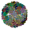
 Sample preparation
 Electron microscopy imaging
 Processing
