Movie
Movie Controller

[English] 日本語
 Yorodumi
- PDB-4blf: Variable internal flexibility characterizes the helical capsid fo... -
+ Open data

- Basic information
| Entry | Database: PDB / ID: 4blf | ||||||
|---|---|---|---|---|---|---|---|
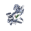
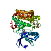
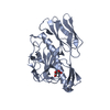
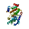
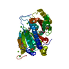
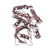
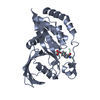
| Title | Variable internal flexibility characterizes the helical capsid formed by Agrobacterium VirE2 protein on single-stranded DNA. | ||||||
 Components Components | SINGLE-STRAND DNA-BINDING PROTEIN | ||||||
 Keywords Keywords | DNA BINDING PROTEIN / TCOMPLEX / AGROBACTERIUM / HELICAL RECONSTRUCTION | ||||||
| Function / homology | VirE2 / VirE2 / DNA-mediated transformation / host cell nucleus / DNA binding / extracellular region / identical protein binding / Single-strand DNA-binding protein Function and homology information Function and homology information | ||||||
| Biological species |  AGROBACTERIUM TUMEFACIENS (bacteria) AGROBACTERIUM TUMEFACIENS (bacteria) | ||||||
| Method | ELECTRON MICROSCOPY / helical reconstruction / cryo EM / Resolution: 20 Å | ||||||
 Authors Authors | Bharat, T.A.M. / Zbaida, D. / Eisenstein, M. / Frankenstein, Z. / Mehlman, T. / Weiner, L. / Sorzano, C.O.S. / Barak, Y. / Albeck, S. / Briggs, J.A.G. ...Bharat, T.A.M. / Zbaida, D. / Eisenstein, M. / Frankenstein, Z. / Mehlman, T. / Weiner, L. / Sorzano, C.O.S. / Barak, Y. / Albeck, S. / Briggs, J.A.G. / Wolf, S.G. / Elbaum, M. | ||||||
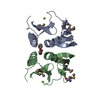
 Citation Citation | Journal: Structure / Year: 2013 Title: Variable internal flexibility characterizes the helical capsid formed by agrobacterium VirE2 protein on single-stranded DNA. Authors: Tanmay A M Bharat / David Zbaida / Miriam Eisenstein / Ziv Frankenstein / Tevie Mehlman / Lev Weiner / Carlos Oscar S Sorzano / Yoav Barak / Shira Albeck / John A G Briggs / Sharon G Wolf / Michael Elbaum /  Abstract: Agrobacterium is known for gene transfer to plants. In addition to a linear ssDNA oligonucleotide, Agrobacterium tumefaciens secretes an abundant ssDNA-binding effector, VirE2. In many ways VirE2 ...Agrobacterium is known for gene transfer to plants. In addition to a linear ssDNA oligonucleotide, Agrobacterium tumefaciens secretes an abundant ssDNA-binding effector, VirE2. In many ways VirE2 adapts the conjugation mechanism to transform the eukaryotic host. The crystal structure of VirE2 shows two compact domains joined by a flexible linker. Bound to ssDNA, VirE2 forms an ordered solenoidal shell, or capsid known as the T-complex. Here, we present a three-dimensional reconstruction of the VirE2-ssDNA complex using cryo-electron microscopy and iterative helical real-space reconstruction. High-resolution refinement was not possible due to inherent heterogeneity in the protein structure. By a combination of computational modeling, chemical modifications, mass spectroscopy, and electron paramagnetic resonance, we found that the N-terminal domain is tightly constrained by both tangential and longitudinal links, while the C terminus is weakly constrained. The quaternary structure is thus rigidly assembled while remaining locally flexible. This flexibility may be important in accommodating substrates without sequence specificity. | ||||||
| History |
|
- Structure visualization
| Movie |
Movie viewer |
|---|---|
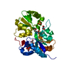
| Structure viewer | Molecule: MolmilJmol/JSmol |
- Downloads & links
-Download
| PDBx/mmCIF format | 4blf.cif.gz | 51.9 KB | Display | PDBx/mmCIF format |
|---|---|---|---|---|
| PDB format | pdb4blf.ent.gz | 37 KB | Display | PDB format |
| PDBx/mmJSON format | 4blf.json.gz | Tree view | PDBx/mmJSON format | |
| Others |  Other downloads Other downloads |
-Validation report
| Arichive directory | https://data.pdbj.org/pub/pdb/validation_reports/bl/4blfftp://data.pdbj.org/pub/pdb/validation_reports/bl/4blf | HTTPS FTP |
|---|
-Related structure data
| Related structure data | 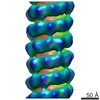 2339MC M: map data used to model this data C: citing same article ( |
|---|---|
| Similar structure data |

-Links
 PDBj
PDBj- Assembly
| Deposited unit | 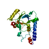
|
|---|---|
| 1 |
|
-Components
| #1: Protein | / 63.5 KDA VIRULENCE PROTEIN / VIRE2 PROTEIN Mass: 26667.943 Da / Num. of mol.: 1 / Fragment: N-TERMINAL DOMAIN, RESIDUES 112-337 Source method: isolated from a genetically manipulated source Source: (gene. exp.) AGROBACTERIUM TUMEFACIENS (bacteria) / Production host: ESCHERICHIA COLI (E. coli) / References: UniProt: P08062 |
|---|
-Experimental details
-Experiment
| Experiment | Method: ELECTRON MICROSCOPY |
|---|---|
| EM experiment | Aggregation state: HELICAL ARRAY / 3D reconstruction method: helical reconstruction |
- Sample preparation
| Component | Name: CRYOEM RECONSTRUCTION OF THE AGROBACTERIUM T-COMPLEX / Type: COMPLEX Details: MICROGRAPHS IN WHICH THON RINGS WERE VISIBLE BEYOND 13 ANGSTROEMS WERE SELECTED. |
|---|---|
| Buffer solution | Name: 50 MM TRIS, 500 MM NACL / pH: 8 / Details: 50 MM TRIS, 500 MM NACL |
| Specimen | Conc.: 1 mg/ml / Embedding applied: NO / Shadowing applied: NO / Staining applied: NO / Vitrification applied: YES |
| Specimen support | Details: HOLEY CARBON |
| Vitrification | Instrument: HOMEMADE PLUNGER / Cryogen name: ETHANE Details: VITRIFICATION 1 -- CRYOGEN- ETHANE, HUMIDITY- 95, INSTRUMENT- HOMEMADE PLUNGER |
- Electron microscopy imaging
| Experimental equipment |  Model: Tecnai F20 / Image courtesy: FEI Company |
|---|---|
| Microscopy | Model: FEI TECNAI F20 / Date: Jun 6, 2008 |
| Electron gun | Electron source: FIELD EMISSION GUN / Accelerating voltage: 200 kV / Illumination mode: FLOOD BEAM |
| Electron lens | Mode: BRIGHT FIELDBright-field microscopy / Nominal magnification: 50000 X / Nominal defocus max: 3200 nm / Nominal defocus min: 1000 nm / Cs: 2 mm |
| Image recording | Electron dose: 20 e/Å2 / Film or detector model: GENERIC TVIPS |
| Radiation wavelength | Relative weight: 1 |
- Processing
| EM software |
| |||||||||||||||||||||
|---|---|---|---|---|---|---|---|---|---|---|---|---|---|---|---|---|---|---|---|---|---|---|
| CTF correction | Details: PHASE-FLIPPING | |||||||||||||||||||||
| 3D reconstruction | Resolution: 20 Å / Num. of particles: 8019 / Nominal pixel size: 4.32 Å / Actual pixel size: 4.32 Å Details: PARTICLES WERE PRE-SELECTED USING XMIPP, AND RECONSTRUCTION WAS CARRIED OUT USING IHRSR PROGRAM IMPLEMENTED IN THE SPIDER PACKAGE. THIS ATOMIC STRUCTURE WAS FITTED INTO THE CRYOEM ENVELOPE ...Details: PARTICLES WERE PRE-SELECTED USING XMIPP, AND RECONSTRUCTION WAS CARRIED OUT USING IHRSR PROGRAM IMPLEMENTED IN THE SPIDER PACKAGE. THIS ATOMIC STRUCTURE WAS FITTED INTO THE CRYOEM ENVELOPE USING FITPDB2EM. SUBMISSION BASED ON EXPERIMENTAL DATA FROM EMDB EMD-2339. (DEPOSITION ID: 11520). Symmetry type: HELICAL | |||||||||||||||||||||
| Atomic model building | Protocol: RIGID BODY FIT / Space: RECIPROCAL / Target criteria: Cross-correlation coefficient / Details: METHOD--RIGID BODY REFINEMENT PROTOCOL--X-RAY | |||||||||||||||||||||
| Atomic model building | PDB-ID: 3BTP | |||||||||||||||||||||
| Refinement | Highest resolution: 20 Å | |||||||||||||||||||||
| Refinement step | Cycle: LAST / Highest resolution: 20 Å
|