- PDB-4bgn: cryo-EM structure of the NavCt voltage-gated sodium channel -
+
Open data
ID or keywords:
Loading...
-
Basic information
Entry
Database: PDB / ID: 4bgn
Title
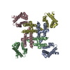
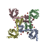
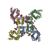
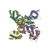
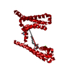
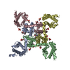
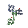
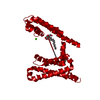
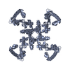
cryo-EM structure of the NavCt voltage-gated sodium channel
Components
VOLTAGE-GATED SODIUM CHANNELSodium channel
Keywords
TRANSPORT PROTEIN
Function / homology
Voltage-dependent channel domain superfamily / Ion transport domain / Ion transport protein / monoatomic ion channel activity / membrane / Ion transport protein
Function and homology information
Biological species
CALDALKALIBACILLUS THERMARUM (bacteria)
Method
ELECTRON CRYSTALLOGRAPHY / electron crystallography / cryo EM / Resolution: 9 Å
Journal: J Mol Biol / Year: 2013 Title: Two alternative conformations of a voltage-gated sodium channel. Authors: Ching-Ju Tsai / Kazutoshi Tani / Katsumasa Irie / Yoko Hiroaki / Takushi Shimomura / Duncan G McMillan / Gregory M Cook / Gebhard F X Schertler / Yoshinori Fujiyoshi / Xiao-Dan Li / Abstract: Activation and inactivation of voltage-gated sodium channels (Navs) are well studied, yet the molecular mechanisms governing channel gating in the membrane remain unknown. We present two ...Activation and inactivation of voltage-gated sodium channels (Navs) are well studied, yet the molecular mechanisms governing channel gating in the membrane remain unknown. We present two conformations of a Nav from Caldalkalibacillus thermarum reconstituted into lipid bilayers in one crystal at 9Å resolution based on electron crystallography. Despite a voltage sensor arrangement identical with that in the activated form, we observed two distinct pore domain structures: a prominent form with a relatively open inner gate and a closed inner-gate conformation similar to the first prokaryotic Nav structure. Structural differences, together with mutational and electrophysiological analyses, indicated that widening of the inner gate was dependent on interactions among the S4-S5 linker, the N-terminal part of S5 and its adjoining part in S6, and on interhelical repulsion by a negatively charged C-terminal region subsequent to S6. Our findings suggest that these specific interactions result in two conformational structures.
Resolution: 9→115 Å / Rfactor Rfree error: 0.058 / Data cutoff high absF: 1042112.63 / Data cutoff low absF: 0 / Isotropic thermal model: RESTRAINED / Cross valid method: THROUGHOUT / σ(F): 0 Details: SUBMISSION BASED ON EXPERIMENTAL DATA FROM EMDB EMD-2347. (DEPOSITION ID: 11571).
In the structure databanks used in Yorodumi, some data are registered as the other names, "COVID-19 virus" and "2019-nCoV". Here are the details of the virus and the list of structure data.
Jan 31, 2019. EMDB accession codes are about to change! (news from PDBe EMDB page)
EMDB accession codes are about to change! (news from PDBe EMDB page)
The allocation of 4 digits for EMDB accession codes will soon come to an end. Whilst these codes will remain in use, new EMDB accession codes will include an additional digit and will expand incrementally as the available range of codes is exhausted. The current 4-digit format prefixed with “EMD-” (i.e. EMD-XXXX) will advance to a 5-digit format (i.e. EMD-XXXXX), and so on. It is currently estimated that the 4-digit codes will be depleted around Spring 2019, at which point the 5-digit format will come into force.
The EM Navigator/Yorodumi systems omit the EMD- prefix.
Related info.:Q: What is EMD? / ID/Accession-code notation in Yorodumi/EM Navigator
Yorodumi is a browser for structure data from EMDB, PDB, SASBDB, etc.
This page is also the successor to EM Navigator detail page, and also detail information page/front-end page for Omokage search.
The word "yorodu" (or yorozu) is an old Japanese word meaning "ten thousand". "mi" (miru) is to see.
Related info.:EMDB / PDB / SASBDB / Comparison of 3 databanks / Yorodumi Search / Aug 31, 2016. New EM Navigator & Yorodumi / Yorodumi Papers / Jmol/JSmol / Function and homology information / Changes in new EM Navigator and Yorodumi
 Movie
Movie Controller


 Open data
 Basic information
 Components
 Sodium channel
  Keywords
 Function and homology information

 Authors
 Citation
 Structure visualization
 Downloads & links
 Other downloads

 PDBj
PDBj
 Assembly
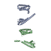

 Sample preparation
 Processing