 Movie
Movie Controller


+ Open data

- Basic information
| Entry | Database: PDB / ID: 3zys | ||||||
|---|---|---|---|---|---|---|---|
| Title | Human dynamin 1 deltaPRD polymer stabilized with GMPPCP | ||||||
 Components Components |
| ||||||
 Keywords Keywords | HYDROLASE/GTP-BINDING PROTEIN / HYDROLASE-GTP-BINDING PROTEIN COMPLEX /  ENDOCYTOSIS / GTP HYDROLYSIS / MEMBRANE REMODELING ENDOCYTOSIS / GTP HYDROLYSIS / MEMBRANE REMODELING | ||||||
| Function / homology |  Function and homology information Function and homology informationclathrin coat assembly involved in endocytosis / vesicle scission / synaptic vesicle budding from presynaptic endocytic zone membrane / presynaptic endocytic zone membrane / interleukin-27-mediated signaling pathway / dynamin GTPase / chromaffin granule / regulation of vesicle size / Toll Like Receptor 4 (TLR4) Cascade / photoreceptor ribbon synapse ...clathrin coat assembly involved in endocytosis / vesicle scission / synaptic vesicle budding from presynaptic endocytic zone membrane / presynaptic endocytic zone membrane / interleukin-27-mediated signaling pathway / dynamin GTPase / chromaffin granule / regulation of vesicle size / Toll Like Receptor 4 (TLR4) Cascade / photoreceptor ribbon synapse / Retrograde neurotrophin signalling / Formation of annular gap junctions / endosome organization / Gap junction degradation / membrane coat / response to type I interferon / negative regulation of viral genome replication / Recycling pathway of L1 / phosphatidylinositol-3,4,5-trisphosphate binding / antiviral innate immune response / synaptic vesicle endocytosis / endocytic vesicle / EPH-ephrin mediated repulsion of cells / clathrin-coated pit / phosphatidylinositol-4,5-bisphosphate binding / MHC class II antigen presentation / receptor-mediated endocytosis / photoreceptor inner segment / cell projection / response to virus / modulation of chemical synaptic transmission / protein homooligomerization / defense response / receptor internalization / ISG15 antiviral mechanism / endocytosis / GDP binding / : / Interferon alpha/beta signaling / presynapse / Clathrin-mediated endocytosis / microtubule binding / protein homotetramerization / defense response to virus / nuclear membrane / microtubule / innate immune response / GTPase activity / glutamatergic synapse / apoptotic process / GTP binding / endoplasmic reticulum membrane / protein kinase binding / perinuclear region of cytoplasm / signal transduction / protein homodimerization activity / RNA binding / extracellular exosome / membrane / identical protein binding / nucleus / plasma membrane / cytosol / cytoplasmSimilarity search - Function | ||||||
| Biological species |  HOMO SAPIENS (human) HOMO SAPIENS (human) | ||||||
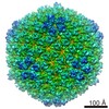
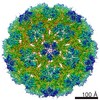
| Method | ELECTRON MICROSCOPY / helical reconstruction / cryo EM / Resolution: 12.2 Å | ||||||
 Authors Authors | Chappie, J.S. / Mears, J.A. / Fang, S. / Leonard, M. / Schmid, S.L. / Milligan, R.A. / Hinshaw, J.E. / Dyda, F. | ||||||
 Citation Citation | Journal: Cell / Year: 2011 Title: A pseudoatomic model of the dynamin polymer identifies a hydrolysis-dependent powerstroke. Authors: Joshua S Chappie / Jason A Mears / Shunming Fang / Marilyn Leonard / Sandra L Schmid / Ronald A Milligan / Jenny E Hinshaw / Fred Dyda /  Abstract: The GTPase dynamin catalyzes membrane fission by forming a collar around the necks of clathrin-coated pits, but the specific structural interactions and conformational changes that drive this process ...The GTPase dynamin catalyzes membrane fission by forming a collar around the necks of clathrin-coated pits, but the specific structural interactions and conformational changes that drive this process remain a mystery. We present the GMPPCP-bound structures of the truncated human dynamin 1 helical polymer at 12.2 Å and a fusion protein, GG, linking human dynamin 1's catalytic G domain to its GTPase effector domain (GED) at 2.2 Å. The structures reveal the position and connectivity of dynamin fragments in the assembled structure, showing that G domain dimers only form between tetramers in sequential rungs of the dynamin helix. Using chemical crosslinking, we demonstrate that dynamin tetramers are made of two dimers, in which the G domain of one molecule interacts in trans with the GED of another. Structural comparison of GG(GMPPCP) to the GG transition-state complex identifies a hydrolysis-dependent powerstroke that may play a role in membrane-remodeling events necessary for fission. | ||||||
| History |
|
- Structure visualization
| Movie |
Movie viewer |
|---|---|
| Structure viewer | Molecule: MolmilJmol/JSmol |
- Downloads & links
-Download
| PDBx/mmCIF format | 3zys.cif.gz | 255.9 KB | Display | PDBx/mmCIF format |
|---|---|---|---|---|
| PDB format | pdb3zys.ent.gz | 197.4 KB | Display | PDB format |
| PDBx/mmJSON format | 3zys.json.gz | Tree view | PDBx/mmJSON format | |
| Others |  Other downloads Other downloads |
-Validation report
| Arichive directory | https://data.pdbj.org/pub/pdb/validation_reports/zy/3zysftp://data.pdbj.org/pub/pdb/validation_reports/zy/3zys | HTTPS FTP |
|---|
-Related structure data
| Related structure data |  1949MC 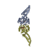 3zycC C: citing same article ( M: map data used to model this data |
|---|---|
| Similar structure data |


-Links
 PDBj
PDBj


- Assembly
| Deposited unit | 
|
|---|---|
| 1 | x 12
|
| 2 |
|
| 3 | 
|
| Symmetry | Helical symmetry: (Circular symmetry: 1 / Dyad axis: no / N subunits divisor: 1 / Num. of operations: 12 / Rise per n subunits: 15.045 Å / Rotation per n subunits: 54.545 °) |
-Components
| #1: Protein | Mass: 39388.961 Da / Num. of mol.: 2 Fragment: G DOMAIN, RESIDUES 1-320, GTPASE EFFECTOR DOMAIN, RESIDUES 726-750 Source method: isolated from a genetically manipulated source Details: EIGHT AMINO ACID POLYPEPTIDE LINKER BETWEEN G DOMAIN AND GTPASE EFFECTOR DOMAIN, RESIDUES 321-328 Source: (gene. exp.) HOMO SAPIENS (human) / Variant: ISOFORM 1 / Plasmid: PMALC2XP5D / Production host:  ESCHERICHIA COLI (E. coli) / Strain (production host): BL21(DE3) / References: UniProt: Q05193, dynamin GTPase ESCHERICHIA COLI (E. coli) / Strain (production host): BL21(DE3) / References: UniProt: Q05193, dynamin GTPase#2: Protein | Mass: 75623.188 Da / Num. of mol.: 2 Source method: isolated from a genetically manipulated source Source: (gene. exp.) HOMO SAPIENS (human) / Plasmid: PSKB-LNB / Production host: ESCHERICHIA COLI (E. coli) / Strain (production host): BL21(DE3) / Variant (production host): ROSETTA / References: UniProt: P20591#3: Protein | Mass: 13440.362 Da / Num. of mol.: 2 / Fragment: PLECKSTRIN HOMOLOGY DOMAIN, RESIDUES 518-630 Source method: isolated from a genetically manipulated source Source: (gene. exp.) HOMO SAPIENS (human) / Production host: ESCHERICHIA COLI (E. coli) / References: UniProt: Q05193, dynamin GTPase |
|---|
-Experimental details
-Experiment
| Experiment | Method: ELECTRON MICROSCOPY |
|---|---|
| EM experiment | Aggregation state: HELICAL ARRAY / 3D reconstruction method: helical reconstruction |
- Sample preparation
| Component | Name: GMPPCP STABILIZED HUMAN DYNAMIN 1 DELTA PRD HELICAL POLYMER Type: COMPLEX Details: DELTA PRD DYNAMIN HELICAL TUBES GENERATED IN THE PRESENCE OF GMPPCP AND DOPS LIPOSOMES |
|---|---|
| Buffer solution | pH: 7.5 |
| Specimen | Embedding applied: NO / Shadowing applied: NO / Staining applied: NO / Vitrification applied: YES |
| Specimen support | Details: HOLEY CARBON |
| Vitrification | Instrument: HOMEMADE PLUNGER / Cryogen name: ETHANE / Details: PLUNGE FROZEN IN LIQUID ETHANE |
- Electron microscopy imaging
| Experimental equipment |  Model: Tecnai F20 / Image courtesy: FEI Company |
|---|---|
| Microscopy | Model: FEI TECNAI F20 |
| Electron gun | Electron source: FIELD EMISSION GUN / Accelerating voltage: 120 kV / Illumination mode: FLOOD BEAM |
| Electron lens | Mode: BRIGHT FIELDBright-field microscopy / Nominal magnification: 50000 X / Nominal defocus max: 2000 nm / Nominal defocus min: 500 nm |
| Image recording | Electron dose: 10 e/Å2 / Film or detector model: GATAN ULTRASCAN 4000 (4k x 4k) |
| Radiation wavelength | Relative weight: 1 |
- Processing
| EM software |
| ||||||||||||
|---|---|---|---|---|---|---|---|---|---|---|---|---|---|
| CTF correction | Details: INDIVIDUAL IMAGES USING ACE2 | ||||||||||||
| 3D reconstruction | Method: IHRSR / Resolution: 12.2 Å / Num. of particles: 4814 / Nominal pixel size: 2.26 Å Details: THIS MODEL INCORPORATES COORDINATES FROM 3ZYC AND 1DYN, WHICH WERE DOCKED INTO A HELICAL CRYO-EM DENSITY. CHAINS A AND D OF THE MODEL SHOW RESIDUES 6-311 OF HUMAN DYNAMIN 1'S G DOMAIN AND ...Details: THIS MODEL INCORPORATES COORDINATES FROM 3ZYC AND 1DYN, WHICH WERE DOCKED INTO A HELICAL CRYO-EM DENSITY. CHAINS A AND D OF THE MODEL SHOW RESIDUES 6-311 OF HUMAN DYNAMIN 1'S G DOMAIN AND RESIDUES 726-748 OF ITS GTPASE EFFECTOR DOMAIN. CHAINS C AND F INCLUDE RESIDUES 518-630 OF DYNAMIN'S PH DOMAIN. RESIDUE 5 SER IN CHAINS A AND D IS A CLONING ARTIFACT THAT WAS VISIBLE IN THE CRYSTAL STRUCTURE OF 3ZYC. INCORPORATES COORDINATES FROM 3LJB, WHICH WERE DOCKED INTO A HELICAL CRYO-EM DENSITY. CHAINS B AND E INCLUDE RESIDUES 367-435 AND 451-531 OF HUMAN MXA'S MIDDLE DOMAIN AND RESIDUES 576-636 OF IF GTPASE EFFECTOR DOMAIN. SUBMISSION BASED ON EXPERIMENTAL DATA FROM EMDB EMD-1949. (DEPOSITION ID: 10194). Symmetry type: HELICAL | ||||||||||||
| Atomic model building | Protocol: FLEXIBLE FIT / Space: REAL / Details: REFINEMENT PROTOCOL--YUP ALGORITHM | ||||||||||||
| Atomic model building |
| ||||||||||||
| Refinement | Highest resolution: 12.2 Å | ||||||||||||
| Refinement step | Cycle: LAST / Highest resolution: 12.2 Å
|

