 Movie
Movie Controller

[English] 日本語
 Yorodumi
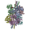
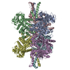
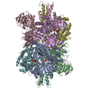
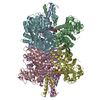
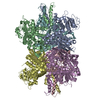
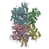
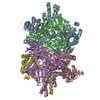
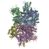
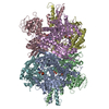
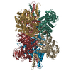
- PDB-3jd3: Glutamate dehydrogenase in complex with NADH and GTP, open confor... -
+ Open data

- Basic information
| Entry | Database: PDB / ID: 3jd3 | ||||||
|---|---|---|---|---|---|---|---|
| Title | Glutamate dehydrogenase in complex with NADH and GTP, open conformation | ||||||
 Components Components | Glutamate dehydrogenase 1, mitochondrial | ||||||
 Keywords Keywords | OXIDOREDUCTASE / glutamate metabolism / mitochondria | ||||||
| Function / homology |  Function and homology information Function and homology informationglutamate dehydrogenase [NAD(P)+] activity / glutamate catabolic process / tricarboxylic acid metabolic process / glutamate dehydrogenase [NAD(P)+] / glutamate dehydrogenase (NADP+) activity / glutamate dehydrogenase (NAD+) activity / glutamine metabolic process / mitochondrial inner membrane / GTP binding / endoplasmic reticulum ...glutamate dehydrogenase [NAD(P)+] activity / glutamate catabolic process / tricarboxylic acid metabolic process / glutamate dehydrogenase [NAD(P)+] / glutamate dehydrogenase (NADP+) activity / glutamate dehydrogenase (NAD+) activity / glutamine metabolic process / mitochondrial inner membrane / GTP binding / endoplasmic reticulum / mitochondrion / ATP binding / identical protein bindingSimilarity search - Function | ||||||
| Biological species |  Bos taurus (cattle) Bos taurus (cattle) | ||||||
| Method | ELECTRON MICROSCOPY / single particle reconstruction / cryo EM / Resolution: 3.6 Å | ||||||
 Authors Authors | Borgnia, M.J. / Banerjee, S. / Merk, A. / Matthies, D. / Bartesaghi, A. / Rao, P. / Pierson, J. / Earl, L.A. / Falconieri, V. / Subramaniam, S. / Milne, J.L.S. | ||||||
 Citation Citation | Journal: Mol Pharmacol / Year: 2016 Title: Using Cryo-EM to Map Small Ligands on Dynamic Metabolic Enzymes: Studies with Glutamate Dehydrogenase. Authors: Mario J Borgnia / Soojay Banerjee / Alan Merk / Doreen Matthies / Alberto Bartesaghi / Prashant Rao / Jason Pierson / Lesley A Earl / Veronica Falconieri / Sriram Subramaniam / Jacqueline L S Milne /  Abstract: Cryo-electron microscopy (cryo-EM) methods are now being used to determine structures at near-atomic resolution and have great promise in molecular pharmacology, especially in the context of mapping ...Cryo-electron microscopy (cryo-EM) methods are now being used to determine structures at near-atomic resolution and have great promise in molecular pharmacology, especially in the context of mapping the binding of small-molecule ligands to protein complexes that display conformational flexibility. We illustrate this here using glutamate dehydrogenase (GDH), a 336-kDa metabolic enzyme that catalyzes the oxidative deamination of glutamate. Dysregulation of GDH leads to a variety of metabolic and neurologic disorders. Here, we report near-atomic resolution cryo-EM structures, at resolutions ranging from 3.2 Å to 3.6 Å for GDH complexes, including complexes for which crystal structures are not available. We show that the binding of the coenzyme NADH alone or in concert with GTP results in a binary mixture in which the enzyme is in either an "open" or "closed" state. Whereas the structure of NADH in the active site is similar between the open and closed states, it is unexpectedly different at the regulatory site. Our studies thus demonstrate that even in instances when there is considerable structural information available from X-ray crystallography, cryo-EM methods can provide useful complementary insights into regulatory mechanisms for dynamic protein complexes. | ||||||
| History |
|
- Structure visualization
| Movie |
Movie viewer |
|---|---|
| Structure viewer | Molecule: MolmilJmol/JSmol |
- Downloads & links
-Download
| PDBx/mmCIF format | 3jd3.cif.gz | 555.8 KB | Display | PDBx/mmCIF format |
|---|---|---|---|---|
| PDB format | pdb3jd3.ent.gz | 461.6 KB | Display | PDB format |
| PDBx/mmJSON format | 3jd3.json.gz | Tree view | PDBx/mmJSON format | |
| Others |  Other downloads Other downloads |
-Validation report
| Arichive directory | https://data.pdbj.org/pub/pdb/validation_reports/jd/3jd3ftp://data.pdbj.org/pub/pdb/validation_reports/jd/3jd3 | HTTPS FTP |
|---|
-Related structure data
| Related structure data | 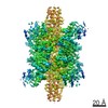 6632MC 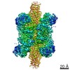 6630C 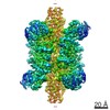 6631C 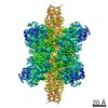 6633C 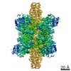 6634C 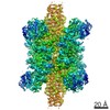 6635C 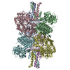 3jczC 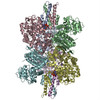 3jd0C  3jd1C 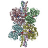 3jd2C 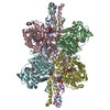 3jd4C M: map data used to model this data C: citing same article ( |
|---|---|
| Similar structure data |
-Links
 PDBj
PDBj

- Assembly
| Deposited unit | 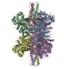
|
|---|---|
| 1 |
|
-Components
| #1: Protein | / GDH 1 Mass: 55802.258 Da / Num. of mol.: 6 / Fragment: UNP residues 58-558 / Source method: isolated from a natural source / Source: (natural) Bos taurus (cattle)References: UniProt: P00366, glutamate dehydrogenase [NAD(P)+] #2: Chemical | ChemComp-GTP / Guanosine triphosphate  Mass: 523.180 Da / Num. of mol.: 6 / Source method: obtained synthetically / Formula: C10H16N5O14P3 / Comment: GTP, energy-carrying molecule*YM Mass: 523.180 Da / Num. of mol.: 6 / Source method: obtained synthetically / Formula: C10H16N5O14P3 / Comment: GTP, energy-carrying molecule*YM#3: Chemical | ChemComp-NAI / Nicotinamide adenine dinucleotide  Mass: 665.441 Da / Num. of mol.: 12 / Source method: obtained synthetically / Formula: C21H29N7O14P2 Mass: 665.441 Da / Num. of mol.: 12 / Source method: obtained synthetically / Formula: C21H29N7O14P2 |
|---|
-Experimental details
-Experiment
| Experiment | Method: ELECTRON MICROSCOPY |
|---|---|
| EM experiment | Aggregation state: PARTICLE / 3D reconstruction method: single particle reconstruction |
- Sample preparation
| Component |
| |||||||||||||||
|---|---|---|---|---|---|---|---|---|---|---|---|---|---|---|---|---|
| Molecular weight | Value: 0.347 MDa / Experimental value: NO | |||||||||||||||
| Buffer solution | Name: 100 mM potassium phosphate, 0.1% n-octyl glucopyranoside, 20 mM GTP, 20 mM NADH pH: 6.8 Details: 100 mM potassium phosphate, 0.1% n-octyl glucopyranoside, 20 mM GTP, 20 mM NADH | |||||||||||||||
| Specimen | Conc.: 2 mg/ml / Embedding applied: NO / Shadowing applied: NO / Staining applied: NO / Vitrification applied: YES | |||||||||||||||
| Specimen support | Details: 200 mesh Quantifoil R2/2 grids (Quantifoil Micro Tools) | |||||||||||||||
| Vitrification | Instrument: FEI VITROBOT MARK IV / Cryogen name: ETHANE / Temp: 90 K / Humidity: 90 % Details: Blot for 3-6 seconds before plunging into liquid ethane (FEI VITROBOT MARK IV). Method: blot for 3-6 seconds before plunging |
- Electron microscopy imaging
| Experimental equipment |  Model: Titan Krios / Image courtesy: FEI Company |
|---|---|
| Microscopy | Model: FEI TITAN KRIOS / Date: Jul 27, 2014 |
| Electron gun | Electron source: FIELD EMISSION GUN / Accelerating voltage: 300 kV / Illumination mode: FLOOD BEAM |
| Electron lens | Mode: BRIGHT FIELDBright-field microscopy / Calibrated magnification: 78426 X / Nominal defocus max: 5000 nm / Nominal defocus min: 1000 nm / Cs: 2.7 mm |
| Specimen holder | Specimen holder model: FEI TITAN KRIOS AUTOGRID HOLDER |
| Image recording | Electron dose: 45 e/Å2 / Film or detector model: GATAN K2 SUMMIT (4k x 4k) |
| EM imaging optics | Energyfilter name: GIF Quantum / Energyfilter upper: 20 eV / Energyfilter lower: 0 eV |
| Image scans | Num. digital images: 1265 |
- Processing
| EM software |
| ||||||||||||||||||||||||||||||||
|---|---|---|---|---|---|---|---|---|---|---|---|---|---|---|---|---|---|---|---|---|---|---|---|---|---|---|---|---|---|---|---|---|---|
| CTF correction | Details: Each micrograph | ||||||||||||||||||||||||||||||||
| Symmetry | Point symmetry: D3 (2x3 fold dihedral) | ||||||||||||||||||||||||||||||||
| 3D reconstruction | Resolution: 3.6 Å / Resolution method: FSC 0.143 CUT-OFF / Num. of particles: 14793 / Nominal pixel size: 0.63754 Å / Actual pixel size: 0.63754 Å / Details: (Single particle--Applied symmetry: D3) / Symmetry type: POINT | ||||||||||||||||||||||||||||||||
| Atomic model building | Protocol: FLEXIBLE FIT / Space: REAL / Details: REFINEMENT PROTOCOL--flexible | ||||||||||||||||||||||||||||||||
| Atomic model building | PDB-ID: 3MW9 3mw9 Pdb chain-ID: A / Accession code: 3MW9 / Source name: PDB / Type: experimental model | ||||||||||||||||||||||||||||||||
| Refinement step | Cycle: LAST
| ||||||||||||||||||||||||||||||||
| Refine LS restraints |
|
