Movie
Movie Controller

[English] 日本語
 Yorodumi
- PDB-3jau: The cryoEM map of EV71 mature viron in complex with the Fab fragm... -
+ Open data

- Basic information
| Entry | Database: PDB / ID: 3jau | ||||||
|---|---|---|---|---|---|---|---|
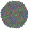
| Title | The cryoEM map of EV71 mature viron in complex with the Fab fragment of antibody D5 | ||||||
 Components Components |
| ||||||
 Keywords Keywords | VIRUS/IMMUNE SYSTEM / Enterovirus 71(EV71) / virus-antibody complex / bivalent binding / high resolution cryo-EM / VIRUS-IMMUNE SYSTEM complex | ||||||
| Function / homology |  Function and homology information Function and homology informationRNA-protein covalent cross-linking / : / : / symbiont-mediated suppression of host cytoplasmic pattern recognition receptor signaling pathway via inhibition of MDA-5 activity / picornain 2A / symbiont-mediated suppression of host mRNA export from nucleus / symbiont genome entry into host cell via pore formation in plasma membrane / picornain 3C / T=pseudo3 icosahedral viral capsid / host cell cytoplasmic vesicle membrane ...RNA-protein covalent cross-linking / : / : / symbiont-mediated suppression of host cytoplasmic pattern recognition receptor signaling pathway via inhibition of MDA-5 activity / picornain 2A / symbiont-mediated suppression of host mRNA export from nucleus / symbiont genome entry into host cell via pore formation in plasma membrane / picornain 3C / T=pseudo3 icosahedral viral capsid / host cell cytoplasmic vesicle membrane / cytoplasmic vesicle membrane / endocytosis involved in viral entry into host cell / symbiont-mediated suppression of host gene expression / viral capsid / nucleoside-triphosphate phosphatase / protein complex oligomerization / monoatomic ion channel activity / RNA helicase activity / symbiont entry into host cell / induction by virus of host autophagy / cysteine-type endopeptidase activity / RNA-directed RNA polymerase / viral RNA genome replication / RNA-dependent RNA polymerase activity / DNA-templated transcription / host cell nucleus / virion attachment to host cell / structural molecule activity / RNA binding / ATP binding / nucleus / metal ion binding Similarity search - Function | ||||||
| Biological species |  Human enterovirus Human enterovirus | ||||||
| Method | ELECTRON MICROSCOPY / single particle reconstruction / cryo EM / Resolution: 4.8 Å | ||||||
 Authors Authors | Fan, C. / Ye, X.H. / Ku, Z.Q. / Zuo, T. / Kong, L.L. / Zhang, C. / Shi, J.P. / Liu, Q.W. / Chen, T. / Zhang, Y.Y. ...Fan, C. / Ye, X.H. / Ku, Z.Q. / Zuo, T. / Kong, L.L. / Zhang, C. / Shi, J.P. / Liu, Q.W. / Chen, T. / Zhang, Y.Y. / Jiang, W. / Zhang, L.Q. / Huang, Z. / Cong, Y. | ||||||
 Citation Citation | Journal: PLoS Pathog / Year: 2016 Title: Structural Basis for Recognition of Human Enterovirus 71 by a Bivalent Broadly Neutralizing Monoclonal Antibody. Authors: Xiaohua Ye / Chen Fan / Zhiqiang Ku / Teng Zuo / Liangliang Kong / Chao Zhang / Jinping Shi / Qingwei Liu / Tan Chen / Yingyi Zhang / Wen Jiang / Linqi Zhang / Zhong Huang / Yao Cong /   Abstract: Enterovirus 71 (EV71) is the main pathogen responsible for hand, foot and mouth disease with severe neurological complications and even death in young children. We have recently identified a highly ...Enterovirus 71 (EV71) is the main pathogen responsible for hand, foot and mouth disease with severe neurological complications and even death in young children. We have recently identified a highly potent anti-EV71 neutralizing monoclonal antibody, termed D5. Here we investigated the structural basis for recognition of EV71 by the antibody D5. Four three-dimensional structures of EV71 particles in complex with IgG or Fab of D5 were reconstructed by cryo-electron microscopy (cryo-EM) single particle analysis all at subnanometer resolutions. The most critical EV71 mature virion-Fab structure was resolved to a resolution of 4.8 Å, which is rare in cryo-EM studies of virus-antibody complex so far. The structures reveal a bivalent binding pattern of D5 antibody across the icosahedral 2-fold axis on mature virion, suggesting that D5 binding may rigidify virions to prevent their conformational changes required for subsequent RNA release. Moreover, we also identified that the complementary determining region 3 (CDR3) of D5 heavy chain directly interacts with the extremely conserved VP1 GH-loop of EV71, which was validated by biochemical and virological assays. We further showed that D5 is indeed able to neutralize a variety of EV71 genotypes and strains. Moreover, D5 could potently confer protection in a mouse model of EV71 infection. Since the conserved VP1 GH-loop is involved in EV71 binding with its uncoating receptor, the scavenger receptor class B, member 2 (SCARB2), the broadly neutralizing ability of D5 might attribute to its inhibition of EV71 from binding SCARB2. Altogether, our results elucidate the structural basis for the binding and neutralization of EV71 by the broadly neutralizing antibody D5, thereby enhancing our understanding of antibody-based protection against EV71 infection. | ||||||
| History |
|
- Structure visualization
| Movie |
Movie viewer |
|---|---|
| Structure viewer | Molecule: MolmilJmol/JSmol |
- Downloads & links
-Download
| PDBx/mmCIF format | 3jau.cif.gz | 51.9 KB | Display | PDBx/mmCIF format |
|---|---|---|---|---|
| PDB format | pdb3jau.ent.gz | 39 KB | Display | PDB format |
| PDBx/mmJSON format | 3jau.json.gz | Tree view | PDBx/mmJSON format | |
| Others |  Other downloads Other downloads |
-Validation report
| Summary document | 3jau_validation.pdf.gz | 924.1 KB | Display | wwPDB validaton report |
|---|---|---|---|---|
| Full document | 3jau_full_validation.pdf.gz | 931.1 KB | Display | |
| Data in XML | 3jau_validation.xml.gz | 17 KB | Display | |
| Data in CIF | 3jau_validation.cif.gz | 22.6 KB | Display | |
| Arichive directory | https://data.pdbj.org/pub/pdb/validation_reports/ja/3jauftp://data.pdbj.org/pub/pdb/validation_reports/ja/3jau | HTTPS FTP |
-Related structure data
| Related structure data | 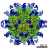 6365MC 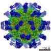 6366MC 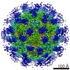 6383MC 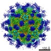 6384MC M: map data used to model this data C: citing same article ( |
|---|---|
| Similar structure data |
-Links
 PDBj
PDBj


- Assembly
| Deposited unit | 
|
|---|---|
| 1 | x 60
|
| 2 |
|
| 3 | x 5
|
| 4 | x 6
|
| 5 | 
|
| Symmetry | Point symmetry: (Schoenflies symbol: I (icosahedral)) |
-Components
| #1: Protein/peptide | Mass: 2001.133 Da / Num. of mol.: 1 / Fragment: UNP residues 207-223 / Source method: isolated from a natural source / Source: (natural) Human enterovirus / References: UniProt: X2L816, UniProt: Q5DW45*PLUS |
|---|---|
| #2: Antibody | Mass: 13026.259 Da / Num. of mol.: 1 / Source method: isolated from a natural source / Source: (natural) Mus musculus |
| #3: Antibody | Mass: 12112.521 Da / Num. of mol.: 1 / Source method: isolated from a natural source / Source: (natural) Mus musculus |
-Experimental details
-Experiment
| Experiment | Method: ELECTRON MICROSCOPY |
|---|---|
| EM experiment | Aggregation state: PARTICLE / 3D reconstruction method: single particle reconstruction |
- Sample preparation
| Component | Name: EV71 mature viron in complex with the Fab fragment of antibody D5 Type: COMPLEX Details: One fab fragment of antibody D5 bind to one protomer of EV71 |
|---|---|
| Molecular weight | Value: 8 MDa / Experimental value: NO |
| Details of virus | Empty: NO / Enveloped: NO / Host category: VERTEBRATES / Isolate: STRAIN / Type: VIRION |
| Natural host | Organism: Homo sapiens |
| Buffer solution | pH: 7.6 / Details: 0.15 M phosphate buffered saline(PBS) buffer |
| Specimen | Embedding applied: NO / Shadowing applied: NO / Staining applied: NO / Vitrification applied: YES Details: 0.15 M phosphate buffered saline(PBS) buffer (Stain Details Grids were plunge-frozen into liquid ethane using a FEI Mark IV virtobot, after 2s blotting to remove extra sample.) |
| Specimen support | Details: 200 mesh R1.2x1.3 Quantifoil Cu grid, glow discharged in air |
| Vitrification | Instrument: FEI VITROBOT MARK IV / Cryogen name: ETHANE / Temp: 120 K / Humidity: 100 % / Method: Blot for 2 seconds before plunging |
- Electron microscopy imaging
| Experimental equipment |  Model: Titan Krios / Image courtesy: FEI Company |
|---|---|
| Microscopy | Model: FEI TITAN KRIOS / Date: Oct 2, 2014 |
| Electron gun | Electron source:  FIELD EMISSION GUN / Accelerating voltage: 300 kV / Illumination mode: FLOOD BEAM FIELD EMISSION GUN / Accelerating voltage: 300 kV / Illumination mode: FLOOD BEAM |
| Electron lens | Mode: BRIGHT FIELD / Nominal magnification: 37000 X / Nominal defocus max: 3500 nm / Nominal defocus min: 2000 nm / Cs: 0.005 mm Astigmatism: Objective lens astigmatism was corrected at 75000 magnification Camera length: 0 mm |
| Specimen holder | Specimen holder model: FEI TITAN KRIOS AUTOGRID HOLDER / Temperature: 91 K / Temperature (max): 92 K / Temperature (min): 90 K / Tilt angle max: 0 ° / Tilt angle min: 0 ° |
| Image recording | Electron dose: 16 e/Å2 / Film or detector model: FEI FALCON II (4k x 4k) |
- Processing
| EM software |
| ||||||||||||
|---|---|---|---|---|---|---|---|---|---|---|---|---|---|
| CTF correction | Details: Each particle | ||||||||||||
| Symmetry | Point symmetry: I (icosahedral) | ||||||||||||
| 3D reconstruction | Method: common lines / Resolution: 4.8 Å / Resolution method: FSC 0.143 CUT-OFF / Num. of particles: 2902 Details: (Single particle details: The particles were boxed using e2boxer.py. CTF fitting was automatically performed using fitctf2.py in jspr, then visually validated and adjusted using EMAN1.9 ...Details: (Single particle details: The particles were boxed using e2boxer.py. CTF fitting was automatically performed using fitctf2.py in jspr, then visually validated and adjusted using EMAN1.9 ctfit program. The gold standard 3D reconstruction procedure was followed using jspr package, with the datasets split into two halves in the beginning.) (Single particle--Applied symmetry: I) Refinement type: HALF-MAPS REFINED INDEPENDENTLY / Symmetry type: POINT | ||||||||||||
| Atomic model building | Protocol: RIGID BODY FIT / Space: REAL / Details: REFINEMENT PROTOCOL--rigid body | ||||||||||||
| Atomic model building | PDB-ID: 3VBS Pdb chain-ID: D | ||||||||||||
| Refinement step | Cycle: LAST
|