 Movie
Movie Controller

[English] 日本語
 Yorodumi
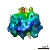
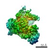
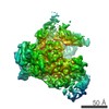
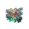
- PDB-3ja7: Cryo-EM structure of the bacteriophage T4 portal protein assembly... -
+ Open data

- Basic information
| Entry | Database: PDB / ID: 3ja7 | ||||||
|---|---|---|---|---|---|---|---|
| Title | Cryo-EM structure of the bacteriophage T4 portal protein assembly at near-atomic resolution | ||||||
 Components Components | Portal protein gp20 | ||||||
 Keywords Keywords |  VIRAL PROTEIN VIRAL PROTEIN | ||||||
| Function / homology | Portal protein Gp20 / Bacteriophage T4-like portal protein (Gp20) / symbiont genome ejection through host cell envelope, contractile tail mechanism / viral portal complex / viral genome packaging / viral release from host cell / host cell plasma membrane / membrane / Portal protein Function and homology information Function and homology information | ||||||
| Biological species |  Enterobacteria phage T4 (virus) Enterobacteria phage T4 (virus) | ||||||
| Method | ELECTRON MICROSCOPY / single particle reconstruction / cryo EM / Resolution: 3.6 Å | ||||||
 Authors Authors | Sun, L. / Zhang, X. / Gao, S. / Rao, P.A. / Padilla-Sanchez, V. / Chen, Z. / Sun, S. / Xiang, Y. / Subramaniam, S. / Rao, V.B. / Rossmann, M.G. | ||||||
 Citation Citation | Journal: Nat Commun / Year: 2015 Title: Cryo-EM structure of the bacteriophage T4 portal protein assembly at near-atomic resolution. Authors: Lei Sun / Xinzheng Zhang / Song Gao / Prashant A Rao / Victor Padilla-Sanchez / Zhenguo Chen / Siyang Sun / Ye Xiang / Sriram Subramaniam / Venigalla B Rao / Michael G Rossmann /  Abstract: The structure and assembly of bacteriophage T4 has been extensively studied. However, the detailed structure of the portal protein remained unknown. Here we report the structure of the bacteriophage ...The structure and assembly of bacteriophage T4 has been extensively studied. However, the detailed structure of the portal protein remained unknown. Here we report the structure of the bacteriophage T4 portal assembly, gene product 20 (gp20), determined by cryo-electron microscopy (cryo-EM) to 3.6 Å resolution. In addition, analysis of a 10 Å resolution cryo-EM map of an empty prolate T4 head shows how the dodecameric portal assembly interacts with the capsid protein gp23 at the special pentameric vertex. The gp20 structure also verifies that the portal assembly is required for initiating head assembly, for attachment of the packaging motor, and for participation in DNA packaging. Comparison of the Myoviridae T4 portal structure with the known portal structures of φ29, SPP1 and P22, representing Podo- and Siphoviridae, shows that the portal structure probably dates back to a time when self-replicating microorganisms were being established on Earth. | ||||||
| History |
|
- Structure visualization
| Movie |
Movie viewer |
|---|---|
| Structure viewer | Molecule: MolmilJmol/JSmol |
- Downloads & links
-Download
| PDBx/mmCIF format | 3ja7.cif.gz | 991.7 KB | Display | PDBx/mmCIF format |
|---|---|---|---|---|
| PDB format | pdb3ja7.ent.gz | 828.8 KB | Display | PDB format |
| PDBx/mmJSON format | 3ja7.json.gz | Tree view | PDBx/mmJSON format | |
| Others |  Other downloads Other downloads |
-Validation report
| Arichive directory | https://data.pdbj.org/pub/pdb/validation_reports/ja/3ja7ftp://data.pdbj.org/pub/pdb/validation_reports/ja/3ja7 | HTTPS FTP |
|---|
-Related structure data
| Related structure data | 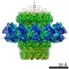 6324MC 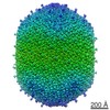 6323C M: map data used to model this data C: citing same article ( |
|---|---|
| Similar structure data |
-Links
 PDBj
PDBj- Assembly
| Deposited unit | 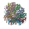
|
|---|---|
| 1 |
|
-Components
| #1: Protein | Mass: 53358.414 Da / Num. of mol.: 12 / Fragment: UNP residues 74-524 Source method: isolated from a genetically manipulated source Source: (gene. exp.) Enterobacteria phage T4 (virus) / Gene: 20 / Plasmid: pCDFDuet-1 / Production host:  Escherichia coli BL21(DE3) (bacteria) / Strain (production host): BL21(DE3) pLysS / References: UniProt: P13334 Escherichia coli BL21(DE3) (bacteria) / Strain (production host): BL21(DE3) pLysS / References: UniProt: P13334 |
|---|
-Experimental details
-Experiment
| Experiment | Method: ELECTRON MICROSCOPY |
|---|---|
| EM experiment | Aggregation state: PARTICLE / 3D reconstruction method: single particle reconstruction |
- Sample preparation
| Component | Name: T4 portal protein gp20 / Type: VIRUS / Details: dodecamer |
|---|---|
| Molecular weight | Value: 0.66 MDa / Experimental value: YES |
| Buffer solution | Name: 20 mM Tris-HCl, 400 mM NaCl, 5 mM EDTA / pH: 7.8 / Details: 20 mM Tris-HCl, 400 mM NaCl, 5 mM EDTA |
| Specimen | Conc.: 1 mg/ml / Embedding applied: NO / Shadowing applied: NO / Staining applied: NO / Vitrification applied: YES |
| Specimen support | Details: Quantifoil R1.2/1.3 holey carbon on 200 mesh copper |
| Vitrification | Instrument: GATAN CRYOPLUNGE 3 / Cryogen name: ETHANE / Temp: 120 K / Humidity: 90 % Details: Blot for 7 seconds before plunging into liquid ethane (GATAN CRYOPLUNGE 3). Method: Blot for 7 seconds before plunging |
- Electron microscopy imaging
| Experimental equipment |  Model: Titan Krios / Image courtesy: FEI Company |
|---|---|
| Microscopy | Model: FEI TITAN KRIOS / Date: Jul 10, 2014 |
| Electron gun | Electron source: FIELD EMISSION GUN / Accelerating voltage: 300 kV / Illumination mode: FLOOD BEAM |
| Electron lens | Mode: BRIGHT FIELDBright-field microscopy / Nominal magnification: 29000 X / Calibrated magnification: 50000 X / Nominal defocus max: 2500 nm / Nominal defocus min: 1500 nm / Cs: 2.7 mm |
| Specimen holder | Specimen holder model: FEI TITAN KRIOS AUTOGRID HOLDER / Specimen holder type: Liquid nitrogen-cooled / Temperature: 100 K / Temperature (max): 105 K / Temperature (min): 80 K |
| Image recording | Electron dose: 2 e/Å2 / Film or detector model: GATAN K2 (4k x 4k) |
| EM imaging optics | Energyfilter name: GIF / Energyfilter upper: 25 eV / Energyfilter lower: 0 eV |
| Image scans | Num. digital images: 839 |
| Radiation | Protocol: SINGLE WAVELENGTH / Monochromatic (M) / Laue (L): M / Scattering type: x-ray |
| Radiation wavelength | Relative weight: 1 |
- Processing
| EM software | Name: RELION / Category: 3D reconstruction | |||||||||||||||||||||||||||||||||||||||||||||||||||||||||||||||||||||||||||||||||||||||||||||||||||||||||
|---|---|---|---|---|---|---|---|---|---|---|---|---|---|---|---|---|---|---|---|---|---|---|---|---|---|---|---|---|---|---|---|---|---|---|---|---|---|---|---|---|---|---|---|---|---|---|---|---|---|---|---|---|---|---|---|---|---|---|---|---|---|---|---|---|---|---|---|---|---|---|---|---|---|---|---|---|---|---|---|---|---|---|---|---|---|---|---|---|---|---|---|---|---|---|---|---|---|---|---|---|---|---|---|---|---|---|
| CTF correction | Details: each particle | |||||||||||||||||||||||||||||||||||||||||||||||||||||||||||||||||||||||||||||||||||||||||||||||||||||||||
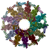
| Symmetry | Point symmetry: C12 (12 fold cyclic) | |||||||||||||||||||||||||||||||||||||||||||||||||||||||||||||||||||||||||||||||||||||||||||||||||||||||||
| 3D reconstruction | Method: Cross-common lines / Resolution: 3.6 Å / Resolution method: FSC 0.143 CUT-OFF / Num. of particles: 98000 / Nominal pixel size: 1 Å / Actual pixel size: 1 Å Details: FSC was calculated from two independent reconstructions. Particles were selected using an automatic selection program. (Single particle--Applied symmetry: C12) Num. of class averages: 15 / Symmetry type: POINT | |||||||||||||||||||||||||||||||||||||||||||||||||||||||||||||||||||||||||||||||||||||||||||||||||||||||||
| Refinement | Resolution: 3.6→3.6 Å / SU ML: 0.48 / σ(F): 0 / Phase error: 28.36 / Stereochemistry target values: ML
| |||||||||||||||||||||||||||||||||||||||||||||||||||||||||||||||||||||||||||||||||||||||||||||||||||||||||
| Solvent computation | Shrinkage radii: 0.9 Å / VDW probe radii: 1.11 Å / Solvent model: FLAT BULK SOLVENT MODEL | |||||||||||||||||||||||||||||||||||||||||||||||||||||||||||||||||||||||||||||||||||||||||||||||||||||||||
| Displacement parameters | Biso max: 302.6 Å2 / Biso mean: 83.0196 Å2 / Biso min: 6.59 Å2 | |||||||||||||||||||||||||||||||||||||||||||||||||||||||||||||||||||||||||||||||||||||||||||||||||||||||||
| Refinement step | Cycle: LAST / Resolution: 3.63→272 Å
| |||||||||||||||||||||||||||||||||||||||||||||||||||||||||||||||||||||||||||||||||||||||||||||||||||||||||
| Refine LS restraints |
| |||||||||||||||||||||||||||||||||||||||||||||||||||||||||||||||||||||||||||||||||||||||||||||||||||||||||
| LS refinement shell | Refine-ID: ELECTRON MICROSCOPY / Total num. of bins used: 14
|
