Movie
Movie Controller

[English] 日本語
 Yorodumi
- PDB-3ja6: Cryo-electron Tomography and All-atom Molecular Dynamics Simulati... -
+ Open data

- Basic information
| Entry | Database: PDB / ID: 3ja6 | ||||||
|---|---|---|---|---|---|---|---|
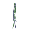
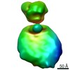
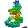
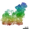
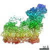
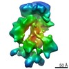
| Title | Cryo-electron Tomography and All-atom Molecular Dynamics Simulations Reveal a Novel Kinase Conformational Switch in Bacterial Chemotaxis Signaling | ||||||
 Components Components |
| ||||||
 Keywords Keywords |  SIGNALING PROTEIN / bacterial chemotaxis / core-signaling unit / adaptor protein / histidine kinase / chemoreceptor SIGNALING PROTEIN / bacterial chemotaxis / core-signaling unit / adaptor protein / histidine kinase / chemoreceptor | ||||||
| Function / homology |  Function and homology informationhistidine kinase / phosphorelay sensor kinase activity / chemotaxis / transmembrane signaling receptor activity / protein domain specific binding / signal transduction / ATP binding / plasma membrane / cytosol / cytoplasm Function and homology informationhistidine kinase / phosphorelay sensor kinase activity / chemotaxis / transmembrane signaling receptor activity / protein domain specific binding / signal transduction / ATP binding / plasma membrane / cytosol / cytoplasmSimilarity search - Function | ||||||
| Biological species |  Escherichia coli (E. coli) Escherichia coli (E. coli) | ||||||
| Method | ELECTRON MICROSCOPY / electron tomography / cryo EM / Resolution: 12.7 Å | ||||||
 Authors Authors | Cassidy, C.K. / Himes, B.A. / Alvarez, F.J. / Ma, J. / Zhao, G. / Perilla, J.R. / Schulten, K. / Zhang, P. | ||||||
 Citation Citation | Journal: Elife / Year: 2015 Title: CryoEM and computer simulations reveal a novel kinase conformational switch in bacterial chemotaxis signaling. Authors: C Keith Cassidy / Benjamin A Himes / Frances J Alvarez / Jun Ma / Gongpu Zhao / Juan R Perilla / Klaus Schulten / Peijun Zhang /  Abstract: Chemotactic responses in bacteria require large, highly ordered arrays of sensory proteins to mediate the signal transduction that ultimately controls cell motility. A mechanistic understanding of ...Chemotactic responses in bacteria require large, highly ordered arrays of sensory proteins to mediate the signal transduction that ultimately controls cell motility. A mechanistic understanding of the molecular events underlying signaling, however, has been hampered by the lack of a high-resolution structural description of the extended array. Here, we report a novel reconstitution of the array, involving the receptor signaling domain, histidine kinase CheA, and adaptor protein CheW, as well as a density map of the core-signaling unit at 11.3 Å resolution, obtained by cryo-electron tomography and sub-tomogram averaging. Extracting key structural constraints from our density map, we computationally construct and refine an atomic model of the core array structure, exposing novel interfaces between the component proteins. Using all-atom molecular dynamics simulations, we further reveal a distinctive conformational change in CheA. Mutagenesis and chemical cross-linking experiments confirm the importance of the conformational dynamics of CheA for chemotactic function. | ||||||
| History |
|
- Structure visualization
| Movie |
Movie viewer |
|---|---|
| Structure viewer | Molecule: MolmilJmol/JSmol |
- Downloads & links
-Download
| PDBx/mmCIF format | 3ja6.cif.gz | 1.5 MB | Display | PDBx/mmCIF format |
|---|---|---|---|---|
| PDB format | pdb3ja6.ent.gz | 1.2 MB | Display | PDB format |
| PDBx/mmJSON format | 3ja6.json.gz | Tree view | PDBx/mmJSON format | |
| Others |  Other downloads Other downloads |
-Validation report
| Arichive directory | https://data.pdbj.org/pub/pdb/validation_reports/ja/3ja6ftp://data.pdbj.org/pub/pdb/validation_reports/ja/3ja6 | HTTPS FTP |
|---|
-Related structure data
| Related structure data | 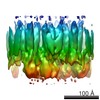 6319MC 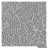 3234C 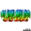 6320C M: map data used to model this data C: citing same article ( |
|---|---|
| Similar structure data |

-Links
 PDBj
PDBj


- Assembly
| Deposited unit | 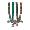
|
|---|---|
| 1 |
|
-Components
| #1: Protein | Mass: 15718.318 Da / Num. of mol.: 4 / Fragment: SEE REMARK 999 Source method: isolated from a genetically manipulated source Source: (gene. exp.) Escherichia coli (E. coli) / Plasmid: PPA770 / Production host: Escherichia coli (E. coli) / Strain (production host): RP3098 / References: UniProt: Q56311*PLUS#2: Protein | Mass: 42489.328 Da / Num. of mol.: 2 Fragment: dimerization domain, kinase domain, and regulatory domain (SEE REMARK 999) Source method: isolated from a genetically manipulated source Source: (gene. exp.) Escherichia coli (E. coli) / Plasmid: pKJ9 / Production host: Escherichia coli (E. coli) / Strain (production host): RP3098 / References: UniProt: Q56310*PLUS#3: Protein | Mass: 33398.828 Da / Num. of mol.: 6 / Fragment: cytoplasmic domain (SEE REMARK 999) Source method: isolated from a genetically manipulated source Source: (gene. exp.) Escherichia coli (E. coli) / Plasmid: pHTCF / Production host: Escherichia coli (E. coli) / Strain (production host): RP3098 / References: UniProt: Q9X0M7*PLUS#4: Protein | Mass: 33225.594 Da / Num. of mol.: 6 / Fragment: cytoplasmic domain (SEE REMARK 999) Source method: isolated from a genetically manipulated source Source: (gene. exp.) Escherichia coli (E. coli) / Plasmid: pHTCF / Production host: Escherichia coli (E. coli) / Strain (production host): RP3098 / References: UniProt: Q9X0M7*PLUSSequence details | THE IMAGED PROTEINS WERE FROM ESCHERICHI | |
|---|
-Experimental details
-Experiment
| Experiment | Method: ELECTRON MICROSCOPY |
|---|---|
| EM experiment | Aggregation state: PARTICLE / 3D reconstruction method: electron tomography |
- Sample preparation
| Component |
| |||||||||||||||||||||||||
|---|---|---|---|---|---|---|---|---|---|---|---|---|---|---|---|---|---|---|---|---|---|---|---|---|---|---|
| Molecular weight | Value: 1.66 MDa / Experimental value: NO | |||||||||||||||||||||||||
| Buffer solution | Name: 75 mM Tris-HCl, 100 mM KCl, 5 mM MgCl2 / pH: 7.4 / Details: 75 mM Tris-HCl, 100 mM KCl, 5 mM MgCl2 | |||||||||||||||||||||||||
| Specimen | Embedding applied: NO / Shadowing applied: NO / Staining applied: NO / Vitrification applied: YES | |||||||||||||||||||||||||
| Specimen support | Details: Perforated R2/2 Quantifoil grids precoated with 10 nm fiducial gold beads on the backside of the grid | |||||||||||||||||||||||||
| Vitrification | Instrument: HOMEMADE PLUNGER / Cryogen name: ETHANE Details: Single-sided blotting (to avoid disruption of the monolayer) before plunging into liquid ethane Method: Single-sided blotting to avoid disruption of the monolayer |
- Electron microscopy imaging
| Experimental equipment |  Model: Tecnai Polara / Image courtesy: FEI Company |
|---|---|
| Microscopy | Model: FEI POLARA 300 / Date: Jan 7, 2009 |
| Electron gun | Electron source: FIELD EMISSION GUN / Accelerating voltage: 200 kV / Illumination mode: SPOT SCAN |
| Electron lens | Mode: BRIGHT FIELDBright-field microscopy / Nominal magnification: 39000 X / Calibrated magnification: 49834 X / Nominal defocus max: 8000 nm / Nominal defocus min: 4000 nm / Cs: 2 mm |
| Specimen holder | Specimen holder model: OTHER / Specimen holder type: Polara cartridge / Tilt angle max: 70 ° / Tilt angle min: -70 ° |
| Image recording | Electron dose: 60 e/Å2 / Film or detector model: GATAN ULTRASCAN 4000 (4k x 4k) |
| Image scans | Num. digital images: 60 |
- Processing
| CTF correction | Details: TomoCTF (strip-based periodogram) | ||||||||||||
|---|---|---|---|---|---|---|---|---|---|---|---|---|---|
| Symmetry | Point symmetry: C2 (2 fold cyclic) | ||||||||||||
| 3D reconstruction | Method: SIRTSirte / Resolution: 12.7 Å / Resolution method: FSC 0.143 CUT-OFF / Num. of particles: 4000 / Nominal pixel size: 3.01 Å / Actual pixel size: 3.01 Å / Details: Gold-standard refinement / Refinement type: HALF-MAPS REFINED INDEPENDENTLY / Symmetry type: 2D CRYSTAL | ||||||||||||
| Atomic model building | Protocol: FLEXIBLE FIT / Space: REAL / Details: REFINEMENT PROTOCOL--FLEXIBLE | ||||||||||||
| Atomic model building | PDB-ID: 1QU7 Accession code: 1QU7 / Source name: PDB / Type: experimental model | ||||||||||||
| Refinement step | Cycle: LAST
|