 Movie
Movie Controller

[English] 日本語
 Yorodumi
- PDB-3j9q: Atomic structures of a bactericidal contractile nanotube in its p... -
+ Open data

- Basic information
| Entry | Database: PDB / ID: 3j9q | ||||||
|---|---|---|---|---|---|---|---|
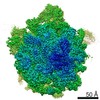
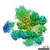
| Title | Atomic structures of a bactericidal contractile nanotube in its pre- and post-contraction states | ||||||
 Components Components |
| ||||||
 Keywords Keywords | STRUCTURAL PROTEIN / pyocin / bacteriocin / sheath / tube | ||||||
| Function / homology |  Function and homology information Function and homology information | ||||||
| Biological species |   Pseudomonas aeruginosa (bacteria) Pseudomonas aeruginosa (bacteria) | ||||||
| Method | ELECTRON MICROSCOPY / helical reconstruction / cryo EM / Resolution: 3.5 Å | ||||||
 Authors Authors | Ge, P. / Scholl, D. / Leiman, P.G. / Yu, X. / Miller, J.F. / Zhou, Z.H. | ||||||
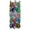
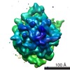
 Citation Citation | Journal: Nat Struct Mol Biol / Year: 2015 Title: Atomic structures of a bactericidal contractile nanotube in its pre- and postcontraction states. Authors: Peng Ge / Dean Scholl / Petr G Leiman / Xuekui Yu / Jeff F Miller / Z Hong Zhou /   Abstract: R-type pyocins are representatives of contractile ejection systems, a class of biological nanomachines that includes, among others, the bacterial type VI secretion system (T6SS) and contractile ...R-type pyocins are representatives of contractile ejection systems, a class of biological nanomachines that includes, among others, the bacterial type VI secretion system (T6SS) and contractile bacteriophage tails. We report atomic models of the Pseudomonas aeruginosa precontraction pyocin sheath and tube, and the postcontraction sheath, obtained by cryo-EM at 3.5-Å and 3.9-Å resolutions, respectively. The central channel of the tube is negatively charged, in contrast to the neutral and positive counterparts in T6SSs and phage tails. The sheath is interwoven by long N- and C-terminal extension arms emanating from each subunit, which create an extensive two-dimensional mesh that has the same connectivity in the extended and contracted state of the sheath. We propose that the contraction process draws energy from electrostatic and shape complementarities to insert the inner tube through bacterial cell membranes to eventually kill the bacteria. | ||||||
| History |
|
- Structure visualization
| Movie |
Movie viewer |
|---|---|
| Structure viewer | Molecule: MolmilJmol/JSmol |
- Downloads & links
-Download
| PDBx/mmCIF format | 3j9q.cif.gz | 2.1 MB | Display | PDBx/mmCIF format |
|---|---|---|---|---|
| PDB format | pdb3j9q.ent.gz | 1.8 MB | Display | PDB format |
| PDBx/mmJSON format | 3j9q.json.gz | Tree view | PDBx/mmJSON format | |
| Others |  Other downloads Other downloads |
-Validation report
| Summary document | 3j9q_validation.pdf.gz | 1 MB | Display | wwPDB validaton report |
|---|---|---|---|---|
| Full document | 3j9q_full_validation.pdf.gz | 1.1 MB | Display | |
| Data in XML | 3j9q_validation.xml.gz | 283.3 KB | Display | |
| Data in CIF | 3j9q_validation.cif.gz | 436 KB | Display | |
| Arichive directory | https://data.pdbj.org/pub/pdb/validation_reports/j9/3j9qftp://data.pdbj.org/pub/pdb/validation_reports/j9/3j9q | HTTPS FTP |
-Related structure data
| Related structure data | 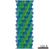 6270MC 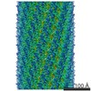 6271C  3j9rC M: map data used to model this data C: citing same article ( |
|---|---|
| Similar structure data |

-Links
 PDBj
PDBj- Assembly
| Deposited unit | 
|
|---|---|
| 1 |
|
| Symmetry | Helical symmetry: (Circular symmetry: 6 / Dyad axis: no / N subunits divisor: 1 / Num. of operations: 3 / Rise per n subunits: 38.4 Å / Rotation per n subunits: 18.3 °) |
-Components
| #1: Protein | Mass: 41247.332 Da / Num. of mol.: 24 / Source method: isolated from a natural source / Source: (natural) Pseudomonas aeruginosa (bacteria) / Strain: PAO / References: UniProt: Q9S574#2: Protein | Mass: 18088.547 Da / Num. of mol.: 24 / Source method: isolated from a natural source / Source: (natural) Pseudomonas aeruginosa (bacteria) / Strain: PAO / References: UniProt: Q9S573 |
|---|
-Experimental details
-Experiment
| Experiment | Method: ELECTRON MICROSCOPY |
|---|---|
| EM experiment | Aggregation state: FILAMENT / 3D reconstruction method: helical reconstruction |
- Sample preparation
| Component | Name: Pyocin (pre-contraction) sheath / Type: COMPLEX / Details: helical |
|---|---|
| Buffer solution | Name: PBS / pH: 7.4 / Details: PBS |
| Specimen | Embedding applied: NO / Shadowing applied: NO / Staining applied: NO / Vitrification applied: YES |
| Specimen support | Details: baked 1.2/1.3 Quantifoil |
| Vitrification | Instrument: FEI VITROBOT MARK IV / Cryogen name: ETHANE / Temp: 90 K / Humidity: 100 % Details: Blot for 4 seconds (blot force 1) before plunging into liquid ethane (FEI VITROBOT MARK IV). Method: Blot time 4 seconds, blot force 1 |
- Electron microscopy imaging
| Experimental equipment |  Model: Titan Krios / Image courtesy: FEI Company |
|---|---|
| Microscopy | Model: FEI TITAN KRIOS / Date: Oct 7, 2009 / Details: Scanned with 9200 ED |
| Electron gun | Electron source:  FIELD EMISSION GUN / Accelerating voltage: 300 kV / Illumination mode: FLOOD BEAM FIELD EMISSION GUN / Accelerating voltage: 300 kV / Illumination mode: FLOOD BEAM |
| Electron lens | Mode: BRIGHT FIELD / Nominal magnification: 59000 X / Nominal defocus max: 2500 nm / Nominal defocus min: 1600 nm / Cs: 2.7 mm |
| Specimen holder | Specimen holder model: FEI TITAN KRIOS AUTOGRID HOLDER / Temperature: 80 K |
| Image recording | Electron dose: 25 e/Å2 / Film or detector model: KODAK SO-163 FILM / Details: Scanned with Nikon 9200 ED |
| Image scans | Num. digital images: 307 |
| Radiation | Protocol: SINGLE WAVELENGTH / Monochromatic (M) / Laue (L): M / Scattering type: x-ray |
| Radiation wavelength | Relative weight: 1 |
- Processing
| EM software |
| ||||||||||||
|---|---|---|---|---|---|---|---|---|---|---|---|---|---|
| CTF correction | Details: Each particle | ||||||||||||
| Helical symmerty | Angular rotation/subunit: 18.3 ° / Axial rise/subunit: 38.4 Å / Axial symmetry: C6 | ||||||||||||
| 3D reconstruction | Method: Relion / Resolution: 3.5 Å / Resolution method: FSC 0.143 CUT-OFF / Nominal pixel size: 1.104 Å / Actual pixel size: 1.104 Å / Details: (Helical Details: Relion-based IHRSR) / Symmetry type: HELICAL | ||||||||||||
| Refinement step | Cycle: LAST
|
