 Movie
Movie Controller


+ Open data

- Basic information
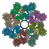
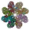
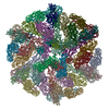
| Entry | Database: PDB / ID: 3j9l | ||||||
|---|---|---|---|---|---|---|---|
| Title | Structure of Dark apoptosome from Drosophila melanogaster | ||||||
 Components Components | Apaf-1 related killer DARK | ||||||
 Keywords Keywords |  APOPTOSIS / programmed cell death APOPTOSIS / programmed cell death | ||||||
| Function / homology |  Function and homology information Function and homology informationnegative regulation of humoral immune response / positive regulation of glial cell apoptotic process / Formation of apoptosome / Regulation of the apoptosome activity / salivary gland histolysis / positive regulation of compound eye retinal cell programmed cell death / melanization defense response / sarcosine catabolic process / central nervous system formation / : ...negative regulation of humoral immune response / positive regulation of glial cell apoptotic process / Formation of apoptosome / Regulation of the apoptosome activity / salivary gland histolysis / positive regulation of compound eye retinal cell programmed cell death / melanization defense response / sarcosine catabolic process / central nervous system formation / : / chaeta development / sperm individualization / apoptosome / S-adenosylmethionine cycle / Neutrophil degranulation / CARD domain binding / programmed cell death / triglyceride homeostasis / dendrite morphogenesis / response to starvation / cysteine-type endopeptidase activator activity involved in apoptotic process / ADP binding / response to gamma radiation / neuron cellular homeostasis / positive regulation of apoptotic process / ATP binding / identical protein bindingSimilarity search - Function | ||||||
| Biological species |  Drosophila melanogaster (fruit fly) Drosophila melanogaster (fruit fly) | ||||||
| Method | ELECTRON MICROSCOPY / single particle reconstruction / cryo EM / Resolution: 4 Å | ||||||
 Authors Authors | Pang, Y. / Bai, X. / Yan, C. / Hao, Q. / Chen, Z. / Wang, J. / Scheres, S.H.W. / Shi, Y. | ||||||
 Citation Citation | Journal: Genes Dev / Year: 2015 Title: Structure of the apoptosome: mechanistic insights into activation of an initiator caspase from Drosophila. Authors: Yuxuan Pang / Xiao-chen Bai / Chuangye Yan / Qi Hao / Zheqin Chen / Jia-Wei Wang / Sjors H W Scheres / Yigong Shi /   Abstract: Apoptosis is executed by a cascade of caspase activation. The autocatalytic activation of an initiator caspase, exemplified by caspase-9 in mammals or its ortholog, Dronc, in fruit flies, is ...Apoptosis is executed by a cascade of caspase activation. The autocatalytic activation of an initiator caspase, exemplified by caspase-9 in mammals or its ortholog, Dronc, in fruit flies, is facilitated by a multimeric adaptor complex known as the apoptosome. The underlying mechanism by which caspase-9 or Dronc is activated by the apoptosome remains unknown. Here we report the electron cryomicroscopic (cryo-EM) structure of the intact apoptosome from Drosophila melanogaster at 4.0 Å resolution. Analysis of the Drosophila apoptosome, which comprises 16 molecules of the Dark protein (Apaf-1 ortholog), reveals molecular determinants that support the assembly of the 2.5-MDa complex. In the absence of dATP or ATP, Dronc zymogen potently induces formation of the Dark apoptosome, within which Dronc is efficiently activated. At 4.1 Å resolution, the cryo-EM structure of the Dark apoptosome bound to the caspase recruitment domain (CARD) of Dronc (Dronc-CARD) reveals two stacked rings of Dronc-CARD that are sandwiched between two octameric rings of the Dark protein. The specific interactions between Dronc-CARD and both the CARD and the WD40 repeats of a nearby Dark protomer are indispensable for Dronc activation. These findings reveal important mechanistic insights into the activation of initiator caspase by the apoptosome. | ||||||
| History |
|
- Structure visualization
| Movie |
Movie viewer |
|---|---|
| Structure viewer | Molecule: MolmilJmol/JSmol |
- Downloads & links
-Download
| PDBx/mmCIF format | 3j9l.cif.gz | 2.6 MB | Display | PDBx/mmCIF format |
|---|---|---|---|---|
| PDB format | pdb3j9l.ent.gz | Display | PDB format | |
| PDBx/mmJSON format | 3j9l.json.gz | Tree view | PDBx/mmJSON format | |
| Others |  Other downloads Other downloads |
-Validation report
| Arichive directory | https://data.pdbj.org/pub/pdb/validation_reports/j9/3j9lftp://data.pdbj.org/pub/pdb/validation_reports/j9/3j9l | HTTPS FTP |
|---|
-Related structure data
| Related structure data | 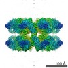 2871MC 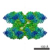 2870C 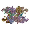 3j9kC M: map data used to model this data C: citing same article ( |
|---|---|
| Similar structure data |


-Links
 PDBj
PDBj


- Assembly
| Deposited unit | 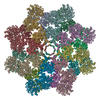
|
|---|---|
| 1 |
|
-Components
| #1: Protein | Mass: 112834.266 Da / Num. of mol.: 16 / Source method: isolated from a natural source / Source: (natural) Drosophila melanogaster (fruit fly) / References: UniProt: Q7KLI1#2: Chemical | ChemComp-DTP / Deoxyadenosine triphosphate  Mass: 491.182 Da / Num. of mol.: 15 / Source method: obtained synthetically / Formula: C10H16N5O12P3 Mass: 491.182 Da / Num. of mol.: 15 / Source method: obtained synthetically / Formula: C10H16N5O12P3 |
|---|
-Experimental details
-Experiment
| Experiment | Method: ELECTRON MICROSCOPY |
|---|---|
| EM experiment | Aggregation state: PARTICLE / 3D reconstruction method: single particle reconstruction |
- Sample preparation
| Component | Name: Dark apoptosome / Type: COMPLEX |
|---|---|
| Molecular weight | Value: 2.5 MDa / Experimental value: NO |
| Buffer solution | Name: 25 mM Tris, pH 8.0, 150 mM NaCl, 5 mM DTT / pH: 8 / Details: 25 mM Tris, pH 8.0, 150 mM NaCl, 5 mM DTT |
| Specimen | Conc.: 0.15 mg/ml / Embedding applied: NO / Shadowing applied: NO / Staining applied: NO / Vitrification applied: YES |
| Specimen support | Details: glow-discharged holey carbon grids (Quantifoil CuR2/2) with home-made continuous carbon film |
| Vitrification | Instrument: FEI VITROBOT MARK II / Cryogen name: ETHANE / Temp: 85 K / Humidity: 100 % Details: Blot for 2 seconds before plunging into liquid ethane (FEI VITROBOT MARK II). Method: Blot for 2 seconds before plunging |
- Electron microscopy imaging
| Experimental equipment |  Model: Tecnai Polara / Image courtesy: FEI Company |
|---|---|
| Microscopy | Model: FEI POLARA 300 / Date: Nov 17, 2013 |
| Electron gun | Electron source: FIELD EMISSION GUN / Accelerating voltage: 300 kV / Illumination mode: FLOOD BEAM |
| Electron lens | Mode: BRIGHT FIELDBright-field microscopy / Nominal magnification: 78000 X / Calibrated magnification: 104748 X / Nominal defocus max: 6600 nm / Nominal defocus min: 1600 nm / Cs: 2 mm |
| Specimen holder | Specimen holder model: GATAN LIQUID NITROGEN / Temperature (max): 90 K / Temperature (min): 80 K |
| Image recording | Electron dose: 28 e/Å2 / Film or detector model: FEI FALCON II (4k x 4k) |
| Image scans | Num. digital images: 693 |
| Radiation | Protocol: SINGLE WAVELENGTH / Monochromatic (M) / Laue (L): M / Scattering type: x-ray |
| Radiation wavelength | Relative weight: 1 |
- Processing
| EM software |
| ||||||||||||
|---|---|---|---|---|---|---|---|---|---|---|---|---|---|
| CTF correction | Details: Each particle | ||||||||||||
| Symmetry | Point symmetry: D8 (2x8 fold dihedral) | ||||||||||||
| 3D reconstruction | Resolution: 4 Å / Resolution method: FSC 0.143 CUT-OFF / Num. of particles: 9354 / Nominal pixel size: 1.34 Å / Actual pixel size: 1.34 Å Details: To correct for beam-induced movements, the 16 video frames for each micrograph were first aligned using whole-image motion correction. Then particle-based beam-induced movement correction ...Details: To correct for beam-induced movements, the 16 video frames for each micrograph were first aligned using whole-image motion correction. Then particle-based beam-induced movement correction was performed using statistical movie processing in RELION. Symmetry type: POINT | ||||||||||||
| Refinement step | Cycle: LAST
|
