[English] 日本語
Yorodumi
- PDB-3j9d: Atomic structure of a non-enveloped virus reveals pH sensors for ... -

+
Open data


ID or keywords:

Loading...

-
Basic information

Entry
Database: PDB / ID: 3j9d
TitleAtomic structure of a non-enveloped virus reveals pH sensors for a coordinated process of cell entry
ComponentsOuter capsid protein VP2
KeywordsVIRAL PROTEIN / non-enveloped virus / cell entry / pH sensor
Function / homologyOuter capsid protein VP2, Orbivirus / Orbivirus outer capsid protein VP2 / clathrin-dependent endocytosis of virus by host cell / structural molecule activity / virion attachment to host cell / Outer capsid protein VP2
Function and homology information
Biological speciesBluetongue virus 1
MethodELECTRON MICROSCOPY / single particle reconstruction / cryo EM / Resolution: 3.3 Å
AuthorsZhang, X. / Patel, A. / Celma, C. / Roy, P. / Zhou, Z.H.
Citation
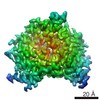
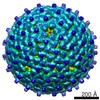
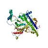
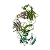
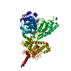
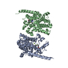
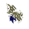
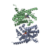
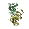
Journal: Nat Struct Mol Biol / Year: 2016
Title: Atomic model of a nonenveloped virus reveals pH sensors for a coordinated process of cell entry.
Authors: Xing Zhang / Avnish Patel / Cristina C Celma / Xuekui Yu / Polly Roy / Z Hong Zhou /
Abstract: Viruses sense environmental cues such as pH to engage in membrane interactions for cell entry during infection, but how nonenveloped viruses sense pH is largely undefined. Here, we report both high- ...Viruses sense environmental cues such as pH to engage in membrane interactions for cell entry during infection, but how nonenveloped viruses sense pH is largely undefined. Here, we report both high- and low-pH structures of bluetongue virus (BTV), which enters cells via a two-stage endosomal process. The receptor-binding protein VP2 possesses a zinc finger that may function to maintain VP2 in a metastable state and a conserved His866, which senses early-endosomal pH. The membrane-penetration protein VP5 has three domains: dagger, unfurling and anchoring. Notably, the β-meander motif of the anchoring domain contains a histidine cluster that can sense late-endosomal pH and also possesses four putative membrane-interaction elements. Exposing BTV to low pH detaches VP2 and dramatically refolds the dagger and unfurling domains of VP5. Our biochemical and structure-guided-mutagenesis studies support these coordinated pH-sensing mechanisms.
History
DepositionJan 9, 2015Deposition site: RCSB / Processing site: RCSB
Revision 1.0Dec 9, 2015Provider: repository / Type: Initial release
Revision 1.1Dec 23, 2015Group: Database references
Revision 1.2Jan 20, 2016Group: Database references
Revision 1.3Jul 18, 2018Group: Data collection / Category: em_software / Item: _em_software.image_processing_id / _em_software.name
Revision 1.4Dec 18, 2019Group: Other / Category: atom_sites
Item: _atom_sites.fract_transf_matrix[1][1] / _atom_sites.fract_transf_matrix[2][2] / _atom_sites.fract_transf_matrix[3][3]
Revision 1.5Feb 21, 2024Group: Data collection / Database references ...Data collection / Database references / Derived calculations / Refinement description
Category: chem_comp_atom / chem_comp_bond ...chem_comp_atom / chem_comp_bond / database_2 / pdbx_struct_conn_angle / refine / struct_conn / struct_site
Item: _database_2.pdbx_DOI / _database_2.pdbx_database_accession ..._database_2.pdbx_DOI / _database_2.pdbx_database_accession / _pdbx_struct_conn_angle.ptnr1_auth_comp_id / _pdbx_struct_conn_angle.ptnr1_auth_seq_id / _pdbx_struct_conn_angle.ptnr1_label_atom_id / _pdbx_struct_conn_angle.ptnr1_label_comp_id / _pdbx_struct_conn_angle.ptnr1_label_seq_id / _pdbx_struct_conn_angle.ptnr3_auth_comp_id / _pdbx_struct_conn_angle.ptnr3_auth_seq_id / _pdbx_struct_conn_angle.ptnr3_label_atom_id / _pdbx_struct_conn_angle.ptnr3_label_comp_id / _pdbx_struct_conn_angle.ptnr3_label_seq_id / _pdbx_struct_conn_angle.value / _refine.ls_d_res_high / _refine.ls_d_res_low / _struct_conn.pdbx_dist_value / _struct_conn.ptnr1_auth_comp_id / _struct_conn.ptnr1_auth_seq_id / _struct_conn.ptnr1_label_atom_id / _struct_conn.ptnr1_label_comp_id / _struct_conn.ptnr1_label_seq_id / _struct_site.pdbx_auth_asym_id / _struct_site.pdbx_auth_comp_id / _struct_site.pdbx_auth_seq_id

-
Structure visualization

Movie
  • Deposited structure unit
  • Imaged by Jmol
  • Download
  • Superimposition on EM map
  • EMDB-6239
  • Imaged by UCSF Chimera
  • Download
Movie viewer
Structure viewerMolecule:
MolmilJmol/JSmol

Downloads & links

-
Assembly

Deposited unit
A: Outer capsid protein VP2
hetero molecules


Theoretical massNumber of molelcules
Total (without water)112,1912
Polymers112,1251
Non-polymers651
Water0
1


  • Idetical with deposited unit
  • defined by author
TypeNameSymmetry operationNumber
identity operation1_555x,y,z1

-
Components

#1: Protein Outer capsid protein VP2 / VP2


Mass: 112125.461 Da / Num. of mol.: 1 / Source method: isolated from a natural source / Source: (natural) Bluetongue virus 1 / References: UniProt: B7U676
#2: Chemical ChemComp-ZN / ZINC ION


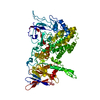
Mass: 65.409 Da / Num. of mol.: 1 / Source method: obtained synthetically / Formula: Zn

-
Experimental details

-
Experiment

ExperimentMethod: ELECTRON MICROSCOPY
EM experimentAggregation state: PARTICLE / 3D reconstruction method: single particle reconstruction

-
Sample preparation

Component
IDNameTypeParent-ID
1Bluetongue Virus 1Bluetongue diseaseVIRUS0
2VP2 of Bluetongue Virus1
Details of virusEmpty: NO / Enveloped: NO / Host category: VERTEBRATES / Isolate: SPECIES / Type: VIRION
Natural hostOrganism: Capra hircus
Buffer solutionpH: 8.8
SpecimenEmbedding applied: NO / Shadowing applied: NO / Staining applied: NO / Vitrification applied: YES
Specimen supportDetails: 400 mesh grid with thin carbon support
VitrificationInstrument: FEI VITROBOT MARK IV / Cryogen name: ETHANE / Temp: 90 K / Humidity: 100 % / Details: Plunged into liquid ethane (FEI VITROBOT MARK IV).

-
Electron microscopy imaging

Experimental equipment
Model: Titan Krios / Image courtesy: FEI Company
MicroscopyModel: FEI TITAN KRIOS / Date: Nov 29, 2013
Electron gunElectron source: FIELD EMISSION GUN / Accelerating voltage: 300 kV / Illumination mode: FLOOD BEAM
Electron lensMode: BRIGHT FIELDBright-field microscopy / Nominal magnification: 14000 X / Calibrated magnification: 24140 X / Nominal defocus max: 3300 nm / Nominal defocus min: 700 nm / Cs: 2.7 mm
Specimen holderSpecimen holder model: FEI TITAN KRIOS AUTOGRID HOLDER / Temperature: 82 K
Image recordingElectron dose: 20 e/Å2 / Film or detector model: GATAN K2 (4k x 4k)
Image scansNum. digital images: 1630

-
Processing

EM softwareName: FREALIGN / Category: 3D reconstruction
CTF correctionDetails: each particle
SymmetryPoint symmetry: I (icosahedral)
3D reconstructionMethod: reference match / Resolution: 3.3 Å / Resolution method: FSC 0.143 CUT-OFF / Num. of particles: 5008 / Nominal pixel size: 1.01 Å / Actual pixel size: 1.01 Å / Symmetry type: POINT
RefinementResolution: 3.3→3.3 Å / SU ML: 0.38 / σ(F): 2 / Phase error: 22.14 / Stereochemistry target values: MLHL
RfactorNum. reflection% reflection
Rfree0.1808 456 0.06 %
Rwork0.1826 784934 -
obs0.1826 785390 99.95 %
Solvent computationShrinkage radii: 0.9 Å / VDW probe radii: 1.11 Å / Solvent model: FLAT BULK SOLVENT MODEL
Displacement parametersBiso max: 243.6 Å2 / Biso mean: 53.5661 Å2 / Biso min: 10.48 Å2
Refinement stepCycle: LAST / Resolution: 3.5→39.924 Å
ProteinNucleic acidLigandSolventTotal
Num. atoms6085 0 1 0 6086
Refine LS restraints
Refine-IDTypeDev idealNumber
ELECTRON MICROSCOPYf_bond_d0.0276221
ELECTRON MICROSCOPYf_angle_d1.9678420
ELECTRON MICROSCOPYf_chiral_restr0.079929
ELECTRON MICROSCOPYf_plane_restr0.0091078
ELECTRON MICROSCOPYf_dihedral_angle_d20.3142342
LS refinement shell

Refine-ID: ELECTRON MICROSCOPY / Total num. of bins used: 3 / % reflection obs: 100 %

Resolution (Å)Rfactor RfreeNum. reflection RfreeRfactor RworkNum. reflection RworkNum. reflection all
3.5-4.00630.30321680.2804261967262135
4.0063-5.04590.13881200.1455261570261690
5.0459-39.92630.14781680.1591261397261565
Refinement TLS params.Method: refined / Origin x: -70.3706 Å / Origin y: 90.8331 Å / Origin z: 351.1993 Å
111213212223313233
T0.2194 Å20.0199 Å2-0.0152 Å2-0.2392 Å20.0014 Å2--0.2144 Å2
L0.6088 °20.3129 °2-0.1275 °2-1.3645 °20.1281 °2--0.7259 °2
S0.0909 Å °-0.1106 Å °0.2265 Å °0.217 Å °-0.032 Å °0.0006 Å °-0.2821 Å °0.047 Å °0.0365 Å °
Refinement TLS groupSelection details: all

+
About Yorodumi

-
News

-
Feb 9, 2022. New format data for meta-information of EMDB entries

New format data for meta-information of EMDB entries

  • Version 3 of the EMDB header file is now the official format.
  • The previous official version 1.9 will be removed from the archive.

Related info.:EMDB header

External links:wwPDB to switch to version 3 of the EMDB data model

-
Aug 12, 2020. Covid-19 info

Covid-19 info

URL: https://pdbj.org/emnavi/covid19.php

New page: Covid-19 featured information page in EM Navigator.

Related info.:Covid-19 info / Mar 5, 2020. Novel coronavirus structure data

+
Mar 5, 2020. Novel coronavirus structure data

Novel coronavirus structure data

Related info.:Yorodumi Speices / Aug 12, 2020. Covid-19 info

External links:COVID-19 featured content - PDBj / Molecule of the Month (242):Coronavirus Proteases

+
Jan 31, 2019. EMDB accession codes are about to change! (news from PDBe EMDB page)

EMDB accession codes are about to change! (news from PDBe EMDB page)

  • The allocation of 4 digits for EMDB accession codes will soon come to an end. Whilst these codes will remain in use, new EMDB accession codes will include an additional digit and will expand incrementally as the available range of codes is exhausted. The current 4-digit format prefixed with “EMD-” (i.e. EMD-XXXX) will advance to a 5-digit format (i.e. EMD-XXXXX), and so on. It is currently estimated that the 4-digit codes will be depleted around Spring 2019, at which point the 5-digit format will come into force.
  • The EM Navigator/Yorodumi systems omit the EMD- prefix.

Related info.:Q: What is EMD? / ID/Accession-code notation in Yorodumi/EM Navigator

External links:EMDB Accession Codes are Changing Soon! / Contact to PDBj

+
Jul 12, 2017. Major update of PDB

Major update of PDB

  • wwPDB released updated PDB data conforming to the new PDBx/mmCIF dictionary.
  • This is a major update changing the version number from 4 to 5, and with Remediation, in which all the entries are updated.
  • In this update, many items about electron microscopy experimental information are reorganized (e.g. em_software).
  • Now, EM Navigator and Yorodumi are based on the updated data.

External links:wwPDB Remediation / Enriched Model Files Conforming to OneDep Data Standards Now Available in the PDB FTP Archive

-
Yorodumi

Thousand views of thousand structures

  • Yorodumi is a browser for structure data from EMDB, PDB, SASBDB, etc.
  • This page is also the successor to EM Navigator detail page, and also detail information page/front-end page for Omokage search.
  • The word "yorodu" (or yorozu) is an old Japanese word meaning "ten thousand". "mi" (miru) is to see.

Related info.:EMDB / PDB / SASBDB / Comparison of 3 databanks / Yorodumi Search / Aug 31, 2016. New EM Navigator & Yorodumi / Yorodumi Papers / Jmol/JSmol / Function and homology information / Changes in new EM Navigator and Yorodumi

Read more