 Movie
Movie Controller


+ Open data

- Basic information
| Entry | Database: PDB / ID: 3j83 | ||||||
|---|---|---|---|---|---|---|---|
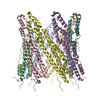
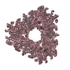
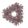
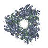
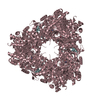
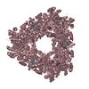
| Title | Heptameric EspB Rosetta model guided by EM density | ||||||
 Components Components | ESX-1 secretion-associated protein EspB | ||||||
 Keywords Keywords |  PROTEIN BINDING PROTEIN BINDING | ||||||
| Function / homology | ESX-1 secretion-associated protein EspB, PE domain / ESX-1 secreted protein B PE domain / protein secretion by the type VII secretion system / PPE superfamily / biological process involved in interaction with host / extracellular region / identical protein binding / ESX-1 secretion-associated protein EspB Function and homology information Function and homology information | ||||||
| Biological species |  Mycobacterium tuberculosis H37Rv (bacteria) Mycobacterium tuberculosis H37Rv (bacteria) | ||||||
| Method | ELECTRON MICROSCOPY / single particle reconstruction / negative staining / Resolution: 30 Å | ||||||
 Authors Authors | Solomonson, M. / DiMaio, F. / Strynadka, N.C.J. | ||||||
 Citation Citation | Journal: Structure / Year: 2015 Title: Structure of EspB from the ESX-1 type VII secretion system and insights into its export mechanism. Authors: Matthew Solomonson / Dheva Setiaputra / Karl A T Makepeace / Emilie Lameignere / Evgeniy V Petrotchenko / Deborah G Conrady / Julien R Bergeron / Marija Vuckovic / Frank DiMaio / Christoph H ...Authors: Matthew Solomonson / Dheva Setiaputra / Karl A T Makepeace / Emilie Lameignere / Evgeniy V Petrotchenko / Deborah G Conrady / Julien R Bergeron / Marija Vuckovic / Frank DiMaio / Christoph H Borchers / Calvin K Yip / Natalie C J Strynadka /   Abstract: Mycobacterium tuberculosis (Mtb) uses the ESX-1 type VII secretion system to export virulence proteins across its lipid-rich cell wall, which helps permeabilize the host's macrophage phagosomal ...Mycobacterium tuberculosis (Mtb) uses the ESX-1 type VII secretion system to export virulence proteins across its lipid-rich cell wall, which helps permeabilize the host's macrophage phagosomal membrane, facilitating the escape and cell-to-cell spread of Mtb. ESX-1 membranolytic activity depends on a set of specialized secreted Esp proteins, the structure and specific roles of which are not currently understood. Here, we report the X-ray and electron microscopic structures of the ESX-1-secreted EspB. We demonstrate that EspB adopts a PE/PPE-like fold that mediates oligomerization with apparent heptameric symmetry, generating a barrel-shaped structure with a central pore that we propose contributes to the macrophage killing functions of EspB. Our structural data also reveal unexpected direct interactions between the EspB bipartite secretion signal sequence elements that form a unified aromatic surface. These findings provide insight into how specialized proteins encoded within the ESX-1 locus are targeted for secretion, and for the first time indicate an oligomerization-dependent role for Esp virulence factors. | ||||||
| History |
|
- Structure visualization
| Movie |
Movie viewer |
|---|---|
| Structure viewer | Molecule: MolmilJmol/JSmol |
- Downloads & links
-Download
| PDBx/mmCIF format | 3j83.cif.gz | 593.7 KB | Display | PDBx/mmCIF format |
|---|---|---|---|---|
| PDB format | pdb3j83.ent.gz | 488 KB | Display | PDB format |
| PDBx/mmJSON format | 3j83.json.gz | Tree view | PDBx/mmJSON format | |
| Others |  Other downloads Other downloads |
-Validation report
| Arichive directory | https://data.pdbj.org/pub/pdb/validation_reports/j8/3j83ftp://data.pdbj.org/pub/pdb/validation_reports/j8/3j83 | HTTPS FTP |
|---|
-Related structure data
| Related structure data |  6120MC  4wj1C 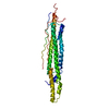 4wj2C M: map data used to model this data C: citing same article ( |
|---|---|
| Similar structure data |
-Links
 PDBj
PDBj- Assembly
| Deposited unit | 
|
|---|---|
| 1 |
|
-Components
| #1: Protein | Mass: 39554.656 Da / Num. of mol.: 7 / Fragment: UNP residues 1-348 Source method: isolated from a genetically manipulated source Source: (gene. exp.) Mycobacterium tuberculosis H37Rv (bacteria)Gene: espB, mtb48, Rv3881c, MTV027.16c / Production host: Escherichia coli (E. coli) / References: UniProt: P9WJD9 |
|---|
-Experimental details
-Experiment
| Experiment | Method: ELECTRON MICROSCOPY |
|---|---|
| EM experiment | Aggregation state: PARTICLE / 3D reconstruction method: single particle reconstruction |
- Sample preparation
| Component | Name: Mycobacterial protein / Type: COMPLEX |
|---|---|
| Molecular weight | Value: 0.23 MDa / Experimental value: YES |
| Buffer solution | Name: 20 mM HEPES, pH 7.5, 150 mM NaCl / pH: 7.5 / Details: 20 mM HEPES, pH 7.5, 150 mM NaCl |
| Specimen | Conc.: 0.01 mg/ml / Embedding applied: NO / Shadowing applied: NO / Staining applied: YES / Vitrification applied: NO Details: Protein was adsorbed onto grid, quickly blotted, and floated on 1% uranyl formate solution for 30 seconds. |
| EM staining | Type: NEGATIVE / Material: uranyl formate |
| Specimen support | Details: 200 mesh gold grid with thin carbon support, glow discharged |
- Electron microscopy imaging
| Experimental equipment |  Model: Tecnai Spirit / Image courtesy: FEI Company |
|---|---|
| Microscopy | Model: FEI TECNAI SPIRIT / Date: Aug 28, 2013 / Details: Low dose |
| Electron gun | Electron source: LAB6 / Accelerating voltage: 120 kV / Illumination mode: FLOOD BEAM |
| Electron lens | Mode: BRIGHT FIELDBright-field microscopy / Nominal magnification: 49000 X / Calibrated magnification: 65900 X / Nominal defocus max: 1200 nm / Nominal defocus min: 800 nm / Camera length: 0 mm |
| Specimen holder | Specimen holder model: SIDE ENTRY, EUCENTRIC Specimen holder type: side entry room temperature tomography holder Temperature: 298 K |
| Image recording | Electron dose: 25 e/Å2 / Film or detector model: FEI EAGLE (4k x 4k) |
| Image scans | Num. digital images: 50 |
- Processing
| EM software |
| ||||||||||||||||
|---|---|---|---|---|---|---|---|---|---|---|---|---|---|---|---|---|---|
| Symmetry | Point symmetry: C7 (7 fold cyclic) | ||||||||||||||||
| 3D reconstruction | Resolution: 30 Å / Resolution method: FSC 0.5 CUT-OFF / Num. of particles: 1455 / Nominal pixel size: 4.7 Å / Actual pixel size: 4.7 Å Details: Initial reconstructions were determined from a side-view 2D average based on observed 7-fold symmetry of the top view using a rotational extrusion method. The initial model was then refined ...Details: Initial reconstructions were determined from a side-view 2D average based on observed 7-fold symmetry of the top view using a rotational extrusion method. The initial model was then refined in 6 iterations with EMAN2 using untilted particles. (Single particle--Applied symmetry: C7) Num. of class averages: 2 / Symmetry type: POINT | ||||||||||||||||
| Atomic model building | Protocol: RIGID BODY FIT / Space: REAL / Target criteria: RMSD / Rosetta score Details: REFINEMENT PROTOCOL--rigid body DETAILS--Homology model generated from PDB coordinates and used for symmetric docking in Rosetta. | ||||||||||||||||
| Atomic model building | PDB-ID: 4WJ1 Pdb chain-ID: A / Accession code: 4WJ1 / Source name: PDB / Type: experimental model | ||||||||||||||||
| Refinement step | Cycle: LAST
|
