[English] 日本語
Yorodumi
- PDB-3j7b: Catalase solved at 3.2 Angstrom resolution by MicroED -

+
Open data


ID or keywords:

Loading...

-
Basic information

Entry
Database: PDB / ID: 3j7b
TitleCatalase solved at 3.2 Angstrom resolution by MicroED
ComponentsCatalase
KeywordsOXIDOREDUCTASE
Function / homology
Function and homology information


Detoxification of Reactive Oxygen Species / catalase complex / cellular detoxification of hydrogen peroxide / Peroxisomal protein import / catalase / catalase activity / Neutrophil degranulation / positive regulation of cell division / hydrogen peroxide catabolic process / response to hydrogen peroxide ...Detoxification of Reactive Oxygen Species / catalase complex / cellular detoxification of hydrogen peroxide / Peroxisomal protein import / catalase / catalase activity / Neutrophil degranulation / positive regulation of cell division / hydrogen peroxide catabolic process / response to hydrogen peroxide / peroxisome / heme binding / enzyme binding / mitochondrion / metal ion binding / cytoplasm
Similarity search - Function
Hemocyanin, N-terminal domain - #60 / Cytochrome C Oxidase; Chain J - #20 / Catalase, clade 3 / Catalase, mono-functional, haem-containing, clades 1 and 3 / Cytochrome C Oxidase; Chain J / Hemocyanin, N-terminal domain / Catalase HpII, Chain A, domain 1 / Catalase core domain / Catalase haem-binding site / Catalase proximal heme-ligand signature. ...Hemocyanin, N-terminal domain - #60 / Cytochrome C Oxidase; Chain J - #20 / Catalase, clade 3 / Catalase, mono-functional, haem-containing, clades 1 and 3 / Cytochrome C Oxidase; Chain J / Hemocyanin, N-terminal domain / Catalase HpII, Chain A, domain 1 / Catalase core domain / Catalase haem-binding site / Catalase proximal heme-ligand signature. / Catalase / Catalase active site / Catalase proximal active site signature. / Catalase immune-responsive domain / Catalase-related immune-responsive / Catalase core domain / Catalase, mono-functional, haem-containing / Catalase / catalase family profile. / Catalase superfamily / Few Secondary Structures / Irregular / Up-down Bundle / Beta Barrel / Mainly Beta / Mainly Alpha
Similarity search - Domain/homology
PROTOPORPHYRIN IX CONTAINING FE / Chem-NDP / Catalase
Similarity search - Component
Biological speciesBos taurus (cattle)
MethodELECTRON CRYSTALLOGRAPHY / electron crystallography / cryo EM / Resolution: 3.2 Å
AuthorsNannenga, B.L. / Shi, D. / Hattne, J. / Reyes, F.E. / Gonen, T.
Citation
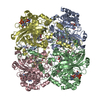
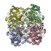
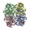
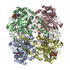
Journal: Elife / Year: 2014
Title: Structure of catalase determined by MicroED.
Authors: Brent L Nannenga / Dan Shi / Johan Hattne / Francis E Reyes / Tamir Gonen /
Abstract: MicroED is a recently developed method that uses electron diffraction for structure determination from very small three-dimensional crystals of biological material. Previously we used a series of ...MicroED is a recently developed method that uses electron diffraction for structure determination from very small three-dimensional crystals of biological material. Previously we used a series of still diffraction patterns to determine the structure of lysozyme at 2.9 Å resolution with MicroED (Shi et al., 2013). Here we present the structure of bovine liver catalase determined from a single crystal at 3.2 Å resolution by MicroED. The data were collected by continuous rotation of the sample under constant exposure and were processed and refined using standard programs for X-ray crystallography. The ability of MicroED to determine the structure of bovine liver catalase, a protein that has long resisted atomic analysis by traditional electron crystallography, demonstrates the potential of this method for structure determination.
History
DepositionJun 9, 2014Deposition site: RCSB / Processing site: RCSB
Revision 1.0Oct 8, 2014Provider: repository / Type: Initial release
Revision 1.1Apr 8, 2015Group: Other
Revision 1.2Aug 24, 2022Group: Data collection / Database references / Derived calculations
Category: citation / citation_author ...citation / citation_author / database_2 / em_image_scans / pdbx_struct_conn_angle / struct_conn / struct_site
Item: _citation.journal_abbrev / _citation.journal_volume ..._citation.journal_abbrev / _citation.journal_volume / _citation.page_first / _citation.page_last / _citation.pdbx_database_id_DOI / _citation.pdbx_database_id_PubMed / _citation.title / _citation_author.identifier_ORCID / _database_2.pdbx_DOI / _database_2.pdbx_database_accession / _pdbx_struct_conn_angle.ptnr1_auth_asym_id / _pdbx_struct_conn_angle.ptnr1_label_asym_id / _pdbx_struct_conn_angle.ptnr2_auth_asym_id / _pdbx_struct_conn_angle.ptnr2_label_asym_id / _pdbx_struct_conn_angle.ptnr3_auth_asym_id / _pdbx_struct_conn_angle.ptnr3_label_asym_id / _pdbx_struct_conn_angle.value / _struct_conn.pdbx_dist_value / _struct_conn.ptnr1_auth_asym_id / _struct_conn.ptnr1_label_asym_id / _struct_conn.ptnr2_auth_asym_id / _struct_conn.ptnr2_label_asym_id / _struct_site.pdbx_auth_asym_id / _struct_site.pdbx_auth_comp_id / _struct_site.pdbx_auth_seq_id
Revision 1.3Feb 21, 2024Group: Data collection / Category: chem_comp_atom / chem_comp_bond

-
Structure visualization

Movie
  • Deposited structure unit
  • Imaged by Jmol
  • Download
  • Superimposition on EM map
  • EMDB-6314
  • Imaged by UCSF Chimera
  • Download
Movie viewer
Structure viewerMolecule:
MolmilJmol/JSmol

Downloads & links

-
Assembly

Deposited unit
A: Catalase
B: Catalase
C: Catalase
D: Catalase
hetero molecules


Theoretical massNumber of molelcules
Total (without water)245,44412
Polymers239,9974
Non-polymers5,4488
Water0
1


  • Idetical with deposited unit
  • defined by author&software
TypeNameSymmetry operationNumber
identity operation1_555x,y,z1
Buried area54220 Å2
ΔGint-309 kcal/mol
Surface area59110 Å2
MethodPISA
Unit cell
Length a, b, c (Å)67.842, 172.081, 182.067
Angle α, β, γ (deg.)90.00, 90.00, 90.00
Int Tables number19
Space group name H-MP212121

-
Components

#1: Protein
Catalase /


Mass: 59999.160 Da / Num. of mol.: 4 / Source method: isolated from a natural source / Source: (natural) Bos taurus (cattle) / References: UniProt: P00432, catalase
#2: Chemical
ChemComp-HEM / PROTOPORPHYRIN IX CONTAINING FE / HEME / Heme B


Mass: 616.487 Da / Num. of mol.: 4 / Source method: obtained synthetically / Formula: C34H32FeN4O4
#3: Chemical
ChemComp-NDP / NADPH DIHYDRO-NICOTINAMIDE-ADENINE-DINUCLEOTIDE PHOSPHATE / Nicotinamide adenine dinucleotide phosphate


Mass: 745.421 Da / Num. of mol.: 4 / Source method: obtained synthetically / Formula: C21H30N7O17P3

-
Experimental details

-
Experiment

ExperimentMethod: ELECTRON CRYSTALLOGRAPHY
EM experimentAggregation state: 3D ARRAY / 3D reconstruction method: electron crystallography

-
Sample preparation

ComponentName: Bovine liver catalase / Type: COMPLEX
Buffer solutionpH: 6.3
SpecimenEmbedding applied: NO / Shadowing applied: NO / Staining applied: NO / Vitrification applied: YES
VitrificationInstrument: FEI VITROBOT MARK IV / Cryogen name: ETHANE

-
Data collection

Experimental equipment
Model: Tecnai F20 / Image courtesy: FEI Company
MicroscopyModel: FEI TECNAI F20 / Date: Jun 15, 2014
Electron gunElectron source: FIELD EMISSION GUN / Accelerating voltage: 200 kV / Illumination mode: OTHER
Electron lensMode: DIFFRACTION
Image recordingElectron dose: 0.1 e/Å2 / Film or detector model: TVIPS TEMCAM-F416 (4k x 4k)
RadiationProtocol: SINGLE WAVELENGTH / Monochromatic (M) / Laue (L): M / Scattering type: electron
Radiation wavelengthRelative weight: 1
ReflectionResolution: 3.2→125.061 Å / Num. all: 36043 / Num. obs: 28101

-
Processing

SoftwareName: REFMAC / Version: 5.8.0075 / Classification: refinement
SymmetryPoint symmetry: D2 (2x2 fold dihedral)
3D reconstructionResolution: 3.2 Å / Resolution method: DIFFRACTION PATTERN/LAYERLINES / Symmetry type: 3D CRYSTAL
RefinementResolution: 3.2→125.06 Å / Cor.coef. Fo:Fc: 0.795 / Cor.coef. Fo:Fc free: 0.745 / Cross valid method: THROUGHOUT / σ(F): 0 / ESU R Free: 0.853 / Stereochemistry target values: MAXIMUM LIKELIHOOD / Details: HYDROGENS HAVE BEEN ADDED IN THE RIDING POSITIONS
RfactorNum. reflection% reflectionSelection details
Rfree0.3079 1369 4.9 %RANDOM
Rwork0.2621 26732 --
obs0.2644 28101 77.97 %-
Solvent computationIon probe radii: 0.8 Å / Shrinkage radii: 0.8 Å / VDW probe radii: 1.2 Å / Solvent model: MASK
Displacement parametersBiso max: 34.26 Å2 / Biso mean: 22.123 Å2 / Biso min: 16.36 Å2
Baniso -1Baniso -2Baniso -3
1--1.18 Å2-0 Å2-0 Å2
2---2.26 Å20 Å2
3---3.44 Å2
Refinement stepCycle: LAST / Resolution: 3.2→125.06 Å
ProteinNucleic acidLigandSolventTotal
Num. atoms16068 0 364 0 16432
Refine LS restraints
Refine-IDTypeDev idealDev ideal targetNumber
ELECTRON CRYSTALLOGRAPHYr_bond_refined_d0.010.01916956
ELECTRON CRYSTALLOGRAPHYr_bond_other_d00.0215384
ELECTRON CRYSTALLOGRAPHYr_angle_refined_deg1.2321.96823124
ELECTRON CRYSTALLOGRAPHYr_angle_other_deg3.501335360
ELECTRON CRYSTALLOGRAPHYr_dihedral_angle_1_deg5.19751992
ELECTRON CRYSTALLOGRAPHYr_dihedral_angle_2_deg33.65123.665884
ELECTRON CRYSTALLOGRAPHYr_dihedral_angle_3_deg14.73152596
ELECTRON CRYSTALLOGRAPHYr_dihedral_angle_4_deg19.80215124
ELECTRON CRYSTALLOGRAPHYr_chiral_restr0.0570.22336
ELECTRON CRYSTALLOGRAPHYr_gen_planes_refined0.010.02119520
ELECTRON CRYSTALLOGRAPHYr_gen_planes_other0.0270.024292
ELECTRON CRYSTALLOGRAPHYr_mcbond_it0.3292.2347980
ELECTRON CRYSTALLOGRAPHYr_mcbond_other0.3292.2347979
ELECTRON CRYSTALLOGRAPHYr_mcangle_it0.513.3519968
LS refinement shellResolution: 3.2→3.283 Å / Total num. of bins used: 20
RfactorNum. reflection% reflection
Rfree0.382 90 -
Rwork0.348 1688 -
all-1778 -
obs--68.6 %

+
About Yorodumi

-
News

-
Feb 9, 2022. New format data for meta-information of EMDB entries

New format data for meta-information of EMDB entries

  • Version 3 of the EMDB header file is now the official format.
  • The previous official version 1.9 will be removed from the archive.

Related info.:EMDB header

External links:wwPDB to switch to version 3 of the EMDB data model

-
Aug 12, 2020. Covid-19 info

Covid-19 info

URL: https://pdbj.org/emnavi/covid19.php

New page: Covid-19 featured information page in EM Navigator.

Related info.:Covid-19 info / Mar 5, 2020. Novel coronavirus structure data

+
Mar 5, 2020. Novel coronavirus structure data

Novel coronavirus structure data

Related info.:Yorodumi Speices / Aug 12, 2020. Covid-19 info

External links:COVID-19 featured content - PDBj / Molecule of the Month (242):Coronavirus Proteases

+
Jan 31, 2019. EMDB accession codes are about to change! (news from PDBe EMDB page)

EMDB accession codes are about to change! (news from PDBe EMDB page)

  • The allocation of 4 digits for EMDB accession codes will soon come to an end. Whilst these codes will remain in use, new EMDB accession codes will include an additional digit and will expand incrementally as the available range of codes is exhausted. The current 4-digit format prefixed with “EMD-” (i.e. EMD-XXXX) will advance to a 5-digit format (i.e. EMD-XXXXX), and so on. It is currently estimated that the 4-digit codes will be depleted around Spring 2019, at which point the 5-digit format will come into force.
  • The EM Navigator/Yorodumi systems omit the EMD- prefix.

Related info.:Q: What is EMD? / ID/Accession-code notation in Yorodumi/EM Navigator

External links:EMDB Accession Codes are Changing Soon! / Contact to PDBj

+
Jul 12, 2017. Major update of PDB

Major update of PDB

  • wwPDB released updated PDB data conforming to the new PDBx/mmCIF dictionary.
  • This is a major update changing the version number from 4 to 5, and with Remediation, in which all the entries are updated.
  • In this update, many items about electron microscopy experimental information are reorganized (e.g. em_software).
  • Now, EM Navigator and Yorodumi are based on the updated data.

External links:wwPDB Remediation / Enriched Model Files Conforming to OneDep Data Standards Now Available in the PDB FTP Archive

-
Yorodumi

Thousand views of thousand structures

  • Yorodumi is a browser for structure data from EMDB, PDB, SASBDB, etc.
  • This page is also the successor to EM Navigator detail page, and also detail information page/front-end page for Omokage search.
  • The word "yorodu" (or yorozu) is an old Japanese word meaning "ten thousand". "mi" (miru) is to see.

Related info.:EMDB / PDB / SASBDB / Comparison of 3 databanks / Yorodumi Search / Aug 31, 2016. New EM Navigator & Yorodumi / Yorodumi Papers / Jmol/JSmol / Function and homology information / Changes in new EM Navigator and Yorodumi

Read more