 Movie
Movie Controller

[English] 日本語
 Yorodumi
- PDB-3j6q: Identification of the active sites in the methyltransferases of a... -
+ Open data

- Basic information
| Entry | Database: PDB / ID: 3j6q | ||||||
|---|---|---|---|---|---|---|---|
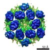
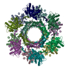
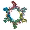
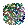
| Title | Identification of the active sites in the methyltransferases of a transcribing dsRNA virus | ||||||
 Components Components | Structural protein VP3 Structure Structure | ||||||
 Keywords Keywords | VIRUS / dsRNA virus / Reoviridae / RNA capping / RNA methyltransferase | ||||||
| Function / homology |  Function and homology information Function and homology information: / : / : / : / Reovirus VP3 protein, guanylyltransferase (GTase) / Reovirus turret protein, bridge domain / Reovirus VP3 protein, Methyltransferase domain 1 / Reovirus VP3 protein, Methyltransferase domain 2 Similarity search - Domain/homology | ||||||
| Biological species |   Bombyx mori cypovirus 1 Bombyx mori cypovirus 1 | ||||||
| Method | ELECTRON MICROSCOPY / single particle reconstruction / cryo EM / Resolution: 3.8 Å | ||||||
 Authors Authors | Zhu, B. / Yang, C. / Liu, H. / Cheng, L. / Song, F. / Zeng, S. / Huang, X. / Ji, G. / Zhu, P. | ||||||
 Citation Citation | Journal: J Mol Biol / Year: 2014 Title: Identification of the active sites in the methyltransferases of a transcribing dsRNA virus. Authors: Bin Zhu / Chongwen Yang / Hongrong Liu / Lingpeng Cheng / Feng Song / Songjun Zeng / Xiaojun Huang / Gang Ji / Ping Zhu /  Abstract: Many double-stranded RNA (dsRNA) viruses are capable of transcribing and capping RNA within a stable icosahedral viral capsid. The turret of turreted dsRNA viruses belonging to the family Reoviridae ...Many double-stranded RNA (dsRNA) viruses are capable of transcribing and capping RNA within a stable icosahedral viral capsid. The turret of turreted dsRNA viruses belonging to the family Reoviridae is formed by five copies of the turret protein, which contains domains with both 7-N-methyltransferase and 2'-O-methyltransferase activities, and serves to catalyze the methylation reactions during RNA capping. Cypovirus of the family Reoviridae provides a good model system for studying the methylation reactions in dsRNA viruses. Here, we present the structure of a transcribing cypovirus to a resolution of ~3.8Å by cryo-electron microscopy. The binding sites for both S-adenosyl-L-methionine and RNA in the two methyltransferases of the turret were identified. Structural analysis of the turret in complex with RNA revealed a pathway through which the RNA molecule reaches the active sites of the two methyltransferases before it is released into the cytoplasm. The pathway shows that RNA capping reactions occur in the active sites of different turret protein monomers, suggesting that RNA capping requires concerted efforts by at least three turret protein monomers. Thus, the turret structure provides novel insights into the precise mechanisms of RNA methylation. | ||||||
| History |
|
- Structure visualization
| Movie |
Movie viewer |
|---|---|
| Structure viewer | Molecule: MolmilJmol/JSmol |
- Downloads & links
-Download
| PDBx/mmCIF format | 3j6q.cif.gz | 903.1 KB | Display | PDBx/mmCIF format |
|---|---|---|---|---|
| PDB format | pdb3j6q.ent.gz | 739.3 KB | Display | PDB format |
| PDBx/mmJSON format | 3j6q.json.gz | Tree view | PDBx/mmJSON format | |
| Others |  Other downloads Other downloads |
-Validation report
| Arichive directory | https://data.pdbj.org/pub/pdb/validation_reports/j6/3j6qftp://data.pdbj.org/pub/pdb/validation_reports/j6/3j6q | HTTPS FTP |
|---|
-Related structure data
| Related structure data |  5926MC M: map data used to model this data C: citing same article ( |
|---|---|
| Similar structure data |
-Links
 PDBj
PDBj- Assembly
| Deposited unit | 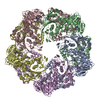
|
|---|---|
| 1 |
|
-Components
| #1: Protein | Structure Mass: 120489.984 Da / Num. of mol.: 5 / Source method: isolated from a natural source / Source: (natural) Bombyx mori cypovirus 1 / References: UniProt: Q914N6#2: Chemical | ChemComp-SAH / S-Adenosyl-L-homocysteine  Type: L-peptide linking / Mass: 384.411 Da / Num. of mol.: 10 / Source method: obtained synthetically / Formula: C14H20N6O5S Type: L-peptide linking / Mass: 384.411 Da / Num. of mol.: 10 / Source method: obtained synthetically / Formula: C14H20N6O5S |
|---|
-Experimental details
-Experiment
| Experiment | Method: ELECTRON MICROSCOPY |
|---|---|
| EM experiment | Aggregation state: PARTICLE / 3D reconstruction method: single particle reconstruction |
- Sample preparation
| Component | Name: transcribing cypovirus / Type: VIRUS |
|---|---|
| Details of virus | Empty: NO / Enveloped: NO / Host category: VERTEBRATES / Isolate: OTHER / Type: VIRION |
| Natural host | Organism: Homo sapiens |
| Specimen | Embedding applied: NO / Shadowing applied: NO / Staining applied: NO / Vitrification applied: YES |
| Vitrification | Instrument: FEI VITROBOT MARK IV / Cryogen name: ETHANE / Humidity: 100 % |
- Electron microscopy imaging
| Experimental equipment |  Model: Titan Krios / Image courtesy: FEI Company |
|---|---|
| Microscopy | Model: FEI TITAN KRIOS / Date: Oct 10, 2012 |
| Electron gun | Electron source: FIELD EMISSION GUN / Accelerating voltage: 300 kV / Illumination mode: OTHER |
| Electron lens | Mode: BRIGHT FIELDBright-field microscopy / Nominal magnification: 75000 X / Calibrated magnification: 125390 X / Nominal defocus max: 3500 nm / Nominal defocus min: 1000 nm / Cs: 2.7 mm / Camera length: 0 mm |
| Specimen holder | Specimen holder model: FEI TITAN KRIOS AUTOGRID HOLDER / Tilt angle max: 0 ° / Tilt angle min: 0 ° |
| Image recording | Film or detector model: GATAN ULTRASCAN 4000 (4k x 4k) |
- Processing
| EM software |
| ||||||||||||
|---|---|---|---|---|---|---|---|---|---|---|---|---|---|
| CTF correction | Details: EMAN | ||||||||||||
| Symmetry | Point symmetry: C1 (asymmetric) | ||||||||||||
| 3D reconstruction | Method: Cross-common lines / Resolution: 3.8 Å / Resolution method: FSC 0.143 CUT-OFF / Num. of particles: 30000 / Details: Applied symmetry: C1 / Symmetry type: POINT | ||||||||||||
| Refinement step | Cycle: LAST
|
