 Movie
Movie Controller

[English] 日本語
 Yorodumi
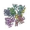
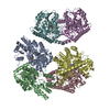
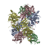
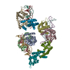
- PDB-3j68: Structural mechanism of the dynein powerstroke (pre-powerstroke state) -
+ Open data

- Basic information
| Entry | Database: PDB / ID: 3j68 | ||||||
|---|---|---|---|---|---|---|---|
| Title | Structural mechanism of the dynein powerstroke (pre-powerstroke state) | ||||||
 Components Components | Dynein motor domain Motor protein Motor protein | ||||||
 Keywords Keywords | MOTOR PROTEIN | ||||||
| Function / homology |  Function and homology informationkaryogamy / establishment of mitotic spindle localization / nuclear migration along microtubule / astral microtubule / minus-end-directed microtubule motor activity / cytoplasmic dynein complex / dynein light intermediate chain binding / spindle pole body / nuclear migration / dynein intermediate chain binding ...karyogamy / establishment of mitotic spindle localization / nuclear migration along microtubule / astral microtubule / minus-end-directed microtubule motor activity / cytoplasmic dynein complex / dynein light intermediate chain binding / spindle pole body / nuclear migration / dynein intermediate chain binding / mitotic sister chromatid segregation / establishment of mitotic spindle orientation / cytoplasmic microtubule / cytoplasmic microtubule organization / Neutrophil degranulation / mitotic spindle organization / cell cortex / ATP hydrolysis activity / ATP binding / cytoplasm Function and homology informationkaryogamy / establishment of mitotic spindle localization / nuclear migration along microtubule / astral microtubule / minus-end-directed microtubule motor activity / cytoplasmic dynein complex / dynein light intermediate chain binding / spindle pole body / nuclear migration / dynein intermediate chain binding ...karyogamy / establishment of mitotic spindle localization / nuclear migration along microtubule / astral microtubule / minus-end-directed microtubule motor activity / cytoplasmic dynein complex / dynein light intermediate chain binding / spindle pole body / nuclear migration / dynein intermediate chain binding / mitotic sister chromatid segregation / establishment of mitotic spindle orientation / cytoplasmic microtubule / cytoplasmic microtubule organization / Neutrophil degranulation / mitotic spindle organization / cell cortex / ATP hydrolysis activity / ATP binding / cytoplasmSimilarity search - Function | ||||||
| Biological species |  Strongylocentrotus purpuratus (purple sea urchin) Strongylocentrotus purpuratus (purple sea urchin) | ||||||
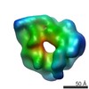
| Method | ELECTRON MICROSCOPY / electron tomography / cryo EM / Resolution: 30 Å | ||||||
 Authors Authors | Lin, J. / Okada, K. / Raytchev, M. / Smith, M.C. / Nicastro, D. | ||||||
 Citation Citation | Journal: Nat Cell Biol / Year: 2014 Title: Structural mechanism of the dynein power stroke. Authors: Jianfeng Lin / Kyoko Okada / Milen Raytchev / Maria C Smith / Daniela Nicastro /  Abstract: Dyneins are large microtubule motor proteins required for mitosis, intracellular transport and ciliary and flagellar motility. They generate force through a power-stroke mechanism, which is an ATP- ...Dyneins are large microtubule motor proteins required for mitosis, intracellular transport and ciliary and flagellar motility. They generate force through a power-stroke mechanism, which is an ATP-consuming cycle of pre- and post-power-stroke conformational changes that cause relative motion between different dynein domains. However, key structural details of dynein's force generation remain elusive. Here, using cryo-electron tomography of intact, active (that is, beating), rapidly frozen sea urchin sperm flagella, we determined the in situ three-dimensional structures of all domains of both pre- and post-power-stroke dynein, including the previously unresolved linker and stalk of pre-power-stroke dynein. Our results reveal that the rotation of the head relative to the linker is the key action in dynein movement, and that there are at least two distinct pre-power-stroke conformations: pre-I (microtubule-detached) and pre-II (microtubule-bound). We provide three-dimensional reconstructions of native dyneins in three conformational states, in situ, allowing us to propose a molecular model of the structural cycle underlying dynein movement. | ||||||
| History |
|
- Structure visualization
| Movie |
Movie viewer |
|---|---|
| Structure viewer | Molecule: MolmilJmol/JSmol |
- Downloads & links
-Download
| PDBx/mmCIF format | 3j68.cif.gz | 464.6 KB | Display | PDBx/mmCIF format |
|---|---|---|---|---|
| PDB format | pdb3j68.ent.gz | 369.5 KB | Display | PDB format |
| PDBx/mmJSON format | 3j68.json.gz | Tree view | PDBx/mmJSON format | |
| Others |  Other downloads Other downloads |
-Validation report
| Arichive directory | https://data.pdbj.org/pub/pdb/validation_reports/j6/3j68ftp://data.pdbj.org/pub/pdb/validation_reports/j6/3j68 | HTTPS FTP |
|---|
-Related structure data
| Related structure data |  5758MC  5757C 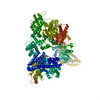 3j67C M: map data used to model this data C: citing same article ( |
|---|---|
| Similar structure data |


-Links
 PDBj
PDBj


- Assembly
| Deposited unit | 
|
|---|---|
| 1 |
|
-Components
| #1: Protein | Motor protein Mass: 262122.234 Da / Num. of mol.: 1 / Fragment: SEE REMARK 999 / Source method: isolated from a natural source Source: (natural) Strongylocentrotus purpuratus (purple sea urchin)References: UniProt: P36022*PLUS |
|---|---|
| Sequence details | THE IMAGED DYNEIN WAS FROM STRONGYLOCENTROTUS PURPURATUS, BUT THE MODELED COORDINATES ARE DERIVED ...THE IMAGED DYNEIN WAS FROM STRONGYLOC |
-Experimental details
-Experiment
| Experiment | Method: ELECTRON MICROSCOPY |
|---|---|
| EM experiment | Aggregation state: PARTICLE / 3D reconstruction method: electron tomography |
- Sample preparation
| Component | Name: active sea urchin sperm flagella / Type: COMPLEX |
|---|---|
| Buffer solution | Name: 360 mM NaCl, 50 mM MgCl2, 10 mM CaCl2, 10 mM KCl, 30 mM HEPES, pH 8.0 pH: 8 Details: 360 mM NaCl, 50 mM MgCl2, 10 mM CaCl2, 10 mM KCl, 30 mM HEPES, pH 8.0 |
| Specimen | Embedding applied: NO / Shadowing applied: NO / Staining applied: NO / Vitrification applied: YES |
| Specimen support | Details: Quantifoil holey carbon grids Cu 200 mesh R2/2 |
| Vitrification | Instrument: HOMEMADE PLUNGER / Cryogen name: ETHANE Details: Blot for 1.5-2.5 seconds before plunging in liquid ethane. Method: Blot for 1.5-2.5 seconds before plunging. |
- Electron microscopy imaging
| Experimental equipment |  Model: Tecnai F30 / Image courtesy: FEI Company |
|---|---|
| Microscopy | Model: FEI TECNAI F30 / Date: Apr 7, 2012 |
| Electron gun | Electron source: FIELD EMISSION GUN / Accelerating voltage: 300 kV / Illumination mode: FLOOD BEAM |
| Electron lens | Mode: BRIGHT FIELDBright-field microscopy / Nominal magnification: 13500 X / Nominal defocus max: 8000 nm / Nominal defocus min: 6000 nm |
| Specimen holder | Specimen holder model: GATAN LIQUID NITROGEN / Specimen holder type: GATAN LIQUID NITROGEN / Temperature: 80 K / Tilt angle max: 65 ° / Tilt angle min: -65 ° |
| Image recording | Electron dose: 100 e/Å2 / Film or detector model: GENERIC GATAN (2k x 2k) |
| EM imaging optics | Energyfilter name: GIF / Energyfilter upper: 20 eV / Energyfilter lower: 0 eV |
| Radiation | Protocol: SINGLE WAVELENGTH / Monochromatic (M) / Laue (L): M / Scattering type: x-ray |
| Radiation wavelength | Relative weight: 1 |
- Processing
| EM software |
| ||||||||||||
|---|---|---|---|---|---|---|---|---|---|---|---|---|---|
| Symmetry | Point symmetry: C1 (asymmetric) | ||||||||||||
| 3D reconstruction | Method: fiducial alignment and weighted back-projection / Resolution: 30 Å / Resolution method: FSC 0.5 CUT-OFF / Nominal pixel size: 9.856 Å / Actual pixel size: 9.856 Å Details: Final maps were calculated by averaging 2800 particles from 41 tomograms. Axonemal repeats (96 nm long) from 41 tomograms (reconstructed using fiducial alignment and weighted backprojection, ...Details: Final maps were calculated by averaging 2800 particles from 41 tomograms. Axonemal repeats (96 nm long) from 41 tomograms (reconstructed using fiducial alignment and weighted backprojection, IMOD software, Kremer et al. 1996) were aligned and averaged using the PEET software (bio3d.colorado.edu, Nicastro et al. 2006). To obtain structures with consistent conformations, classification of the different conformational states of dynein was performed using a clustering approach implemented in PEET (Heumann et al. 2011). Symmetry type: POINT | ||||||||||||
| Atomic model building | Protocol: RIGID BODY FIT / Target criteria: Cross-correlation coefficient Details: REFINEMENT PROTOCOL--rigid body DETAILS--Initial local fitting was done using Chimera and then manual adjustment was performed. | ||||||||||||
| Atomic model building | PDB-ID: 4AKI Pdb chain-ID: A / Accession code: 4AKI / Source name: PDB / Type: experimental model | ||||||||||||
| Refinement step | Cycle: LAST
|
