 Movie
Movie Controller

[English] 日本語
 Yorodumi
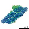
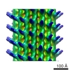
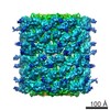
- PDB-3j32: An asymmetric unit map from electron cryo-microscopy of Haliotis ... -
+ Open data

- Basic information
| Entry | Database: PDB / ID: 3j32 | ||||||
|---|---|---|---|---|---|---|---|
| Title | An asymmetric unit map from electron cryo-microscopy of Haliotis diversicolor molluscan hemocyanin isoform 1 (HdH1) | ||||||
 Components Components | Hemocyanin isoform 1 | ||||||
 Keywords Keywords |  OXYGEN TRANSPORT / allosteric OXYGEN TRANSPORT / allosteric | ||||||
| Function / homology |  Function and homology information Function and homology information | ||||||
| Biological species |  Haliotis diversicolor (invertebrata) Haliotis diversicolor (invertebrata) | ||||||
| Method | ELECTRON MICROSCOPY / single particle reconstruction / cryo EM / Resolution: 4.5 Å | ||||||
 Authors Authors | Zhang, Q. / Dai, X. / Cong, Y. / Zhang, J. / Chen, D.-H. / Dougherty, M. / Wang, J. / Ludtke, S. / Schmid, M.F. / Chiu, W. | ||||||
 Citation Citation | Journal: Structure / Year: 2013 Title: Cryo-EM structure of a molluscan hemocyanin suggests its allosteric mechanism. Authors: Qinfen Zhang / Xinghong Dai / Yao Cong / Junjie Zhang / Dong-Hua Chen / Matthew T Dougherty / Jiangyong Wang / Steven J Ludtke / Michael F Schmid / Wah Chiu /  Abstract: Hemocyanins are responsible for transporting O2 in the arthropod and molluscan hemolymph. Haliotis diversicolor molluscan hemocyanin isoform 1 (HdH1) is an 8 MDa oligomer. Each subunit is made up of ...Hemocyanins are responsible for transporting O2 in the arthropod and molluscan hemolymph. Haliotis diversicolor molluscan hemocyanin isoform 1 (HdH1) is an 8 MDa oligomer. Each subunit is made up of eight functional units (FUs). Each FU contains two Cu ions, which can reversibly bind an oxygen molecule. Here, we report a 4.5 A° cryo-EM structure of HdH1. The structure clearly shows ten asymmetric units arranged with D5 symmetry. Each asymmetric unit contains two structurally distinct but chemically identical subunits. The map is sufficiently resolved to trace the entire subunit Ca backbone and to visualize densities corresponding to some large side chains, Cu ion pairs, and interaction networks of adjacent subunits. A FU topology path intertwining between the two subunits of the asymmetric unit is unambiguously determined. Our observations suggest a structural mechanism for the stability of the entire hemocyanin didecamer and 20 ‘‘communication clusters’’ across asymmetric units responsible for its allosteric property upon oxygen binding. | ||||||
| History |
|
- Structure visualization
| Movie |
Movie viewer |
|---|---|
| Structure viewer | Molecule: MolmilJmol/JSmol |
- Downloads & links
-Download
| PDBx/mmCIF format | 3j32.cif.gz | 213.5 KB | Display | PDBx/mmCIF format |
|---|---|---|---|---|
| PDB format | pdb3j32.ent.gz | 122.5 KB | Display | PDB format |
| PDBx/mmJSON format | 3j32.json.gz | Tree view | PDBx/mmJSON format | |
| Others |  Other downloads Other downloads |
-Validation report
| Arichive directory | https://data.pdbj.org/pub/pdb/validation_reports/j3/3j32ftp://data.pdbj.org/pub/pdb/validation_reports/j3/3j32 | HTTPS FTP |
|---|
-Related structure data
-Links
 PDBj
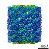
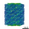
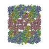
PDBj- Assembly
| Deposited unit | 
|
|---|---|
| 1 | x 10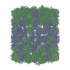
|
| 2 |
|
| 3 | 
|
| Symmetry | Point symmetry: (Schoenflies symbol: D5 (2x5 fold dihedral)) |
-Components
| #1: Protein | Mass: 378672.812 Da / Num. of mol.: 2 / Source method: isolated from a natural source / Source: (natural) Haliotis diversicolor (invertebrata) / Tissue: lymph / References: UniProt: C7FEG7 |
|---|
-Experimental details
-Experiment
| Experiment | Method: ELECTRON MICROSCOPY |
|---|---|
| EM experiment | Aggregation state: PARTICLE / 3D reconstruction method: single particle reconstruction |
- Sample preparation
| Component | Name: An asymmetric unit of Haliotis diversicolor molluscan hemocyanin isoform 1 (HdH1) Type: COMPLEX / Details: one asymmetric unit contains two subunits |
|---|---|
| Molecular weight | Value: 0.8 MDa / Experimental value: NO |
| Buffer solution | Name: 0.2M NaCl, 50mM Tris-HCl, 5mM CaCl2, 5mM MgCl2, pH 7.5 pH: 7.5 Details: 0.2M NaCl, 50mM Tris-HCl, 5mM CaCl2, 5mM MgCl2, pH 7.5 |
| Specimen | Embedding applied: NO / Shadowing applied: NO / Staining applied: NO / Vitrification applied: YES |
| Specimen support | Details: 400 mesh copper 1.2/1.3 quantifoil grid with continuous carbon support, glow discharged. |
| Vitrification | Instrument: FEI VITROBOT MARK III / Cryogen name: ETHANE / Humidity: 100 % Details: Blot for 2 seconds before plunging in to liquid ethane (FEI VITROBOT MARK III) Method: blot for 2 seconds before plunging |
- Electron microscopy imaging
| Microscopy | Model: JEOL 3200FSC / Date: Jul 7, 2008 / Details: MDS |
|---|---|
| Electron gun | Electron source: FIELD EMISSION GUN / Accelerating voltage: 300 kV / Illumination mode: FLOOD BEAM |
| Electron lens | Mode: BRIGHT FIELDBright-field microscopy / Nominal magnification: 60000 X / Nominal defocus max: 3600 nm / Nominal defocus min: 500 nm / Cs: 4.1 mm Astigmatism : Objective lens astigmatism was corrected at 400,000 nominal magnificationCamera length: 0 mm |
| Specimen holder | Specimen holder model: JEOL 3200FSC CRYOHOLDER / Specimen holder type: liquid N2 cooled / Temperature: 88 K / Temperature (max): 100 K / Temperature (min): 80 K / Tilt angle max: 0 ° / Tilt angle min: 0 ° |
| Image recording | Electron dose: 20 e/Å2 / Film or detector model: KODAK SO-163 FILM |
| EM imaging optics | Energyfilter name: In-column Omega Filter / Energyfilter upper: 20 eV / Energyfilter lower: 0 eV |
| Image scans | Num. digital images: 784 |
| Radiation | Protocol: SINGLE WAVELENGTH / Monochromatic (M) / Laue (L): M / Scattering type: x-ray |
| Radiation wavelength | Relative weight: 1 |
- Processing
| EM software | Name: EMAN / Version: 1 / Category: 3D reconstruction | ||||||||||||
|---|---|---|---|---|---|---|---|---|---|---|---|---|---|
| CTF correction | Details: each micrograph | ||||||||||||
| Symmetry | Point symmetry: D5 (2x5 fold dihedral) | ||||||||||||
| 3D reconstruction | Method: projection matching / Resolution: 4.5 Å / Resolution method: FSC 0.5 CUT-OFF / Num. of particles: 28641 / Actual pixel size: 1.02 Å Details: (Single particle details: processed with EMAN1 followed by segmentation of asymmetric unit.) (Single particle--Applied symmetry: D5) Symmetry type: POINT | ||||||||||||
| Refinement step | Cycle: LAST
|
