 Movie
Movie Controller


+ Open data

- Basic information
| Entry | Database: PDB / ID: 3j2v | ||||||
|---|---|---|---|---|---|---|---|
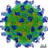
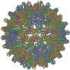
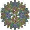
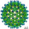
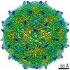
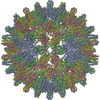
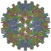
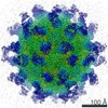
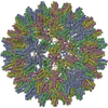
| Title | CryoEM structure of HBV core | ||||||
 Components Components | PreC/core protein | ||||||
 Keywords Keywords | VIRUS / Hepatitis B virus core antigen (HBc) / no Cys61 intermolecular disulfide bond | ||||||
| Function / homology |  Function and homology information Function and homology informationvirus-mediated perturbation of host defense response => GO:0019049 / microtubule-dependent intracellular transport of viral material towards nucleus / T=4 icosahedral viral capsid / : / viral penetration into host nucleus / host cell / host cell cytoplasm / symbiont entry into host cell / structural molecule activity / DNA binding ...virus-mediated perturbation of host defense response => GO:0019049 / microtubule-dependent intracellular transport of viral material towards nucleus / T=4 icosahedral viral capsid / : / viral penetration into host nucleus / host cell / host cell cytoplasm / symbiont entry into host cell / structural molecule activity / DNA binding / RNA binding / extracellular region Similarity search - Function | ||||||
| Biological species |   Hepatitis B virus Hepatitis B virus | ||||||
| Method | ELECTRON MICROSCOPY / single particle reconstruction / cryo EM / Resolution: 3.5 Å | ||||||
 Authors Authors | Yu, X. / Jin, L. / Jih, J. / Shih, C. / Zhou, Z.H. | ||||||
 Citation Citation | Journal: PLoS One / Year: 2013 Title: 3.5Å cryoEM structure of hepatitis B virus core assembled from full-length core protein. Authors: Xuekui Yu / Lei Jin / Jonathan Jih / Chiaho Shih / Z Hong Zhou /  Abstract: The capsid shell of infectious hepatitis B virus (HBV) is composed of 240 copies of a single protein called HBV core antigen (HBc). An atomic model of a core assembled from truncated HBc was ...The capsid shell of infectious hepatitis B virus (HBV) is composed of 240 copies of a single protein called HBV core antigen (HBc). An atomic model of a core assembled from truncated HBc was determined previously by X-ray crystallography. In an attempt to obtain atomic structural information of HBV core in a near native, non-crystalline environment, we reconstructed a 3.5Å-resolution structure of a recombinant core assembled from full-length HBc by cryo electron microscopy (cryoEM) and derived an atomic model. The structure shows that the 240 molecules of full-length HBc form a core with two layers. The outer layer, composed of the N-terminal assembly domain, is similar to the crystal structure of the truncated HBc, but has three differences. First, unlike the crystal structure, our cryoEM structure shows no disulfide bond between the Cys61 residues of the two subunits within the dimer building block, indicating such bond is not required for core formation. Second, our cryoEM structure reveals up to four more residues in the linker region (amino acids 140-149). Third, the loops in the cryoEM structures containing this linker region in subunits B and C are oriented differently (~30° and ~90°) from their counterparts in the crystal structure. The inner layer, composed of the C-terminal arginine-rich domain (ARD) and the ARD-bound RNAs, is partially-ordered and connected with the outer layer through linkers positioned around the two-fold axes. Weak densities emanate from the rims of positively charged channels through the icosahedral three-fold and local three-fold axes. We attribute these densities to the exposed portions of some ARDs, thus explaining ARD's accessibility by proteases and antibodies. Our data supports a role of ARD in mediating communication between inside and outside of the core during HBV maturation and envelopment. | ||||||
| History |
|
- Structure visualization
| Movie |
Movie viewer |
|---|---|
| Structure viewer | Molecule: MolmilJmol/JSmol |
- Downloads & links
-Download
| PDBx/mmCIF format | 3j2v.cif.gz | 125.3 KB | Display | PDBx/mmCIF format |
|---|---|---|---|---|
| PDB format | pdb3j2v.ent.gz | 99.8 KB | Display | PDB format |
| PDBx/mmJSON format | 3j2v.json.gz | Tree view | PDBx/mmJSON format | |
| Others |  Other downloads Other downloads |
-Validation report
| Summary document | 3j2v_validation.pdf.gz | 1.1 MB | Display | wwPDB validaton report |
|---|---|---|---|---|
| Full document | 3j2v_full_validation.pdf.gz | 1.1 MB | Display | |
| Data in XML | 3j2v_validation.xml.gz | 24.4 KB | Display | |
| Data in CIF | 3j2v_validation.cif.gz | 34.5 KB | Display | |
| Arichive directory | https://data.pdbj.org/pub/pdb/validation_reports/j2/3j2vftp://data.pdbj.org/pub/pdb/validation_reports/j2/3j2v | HTTPS FTP |
-Related structure data
| Related structure data | 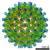 2278MC M: map data used to model this data C: citing same article ( |
|---|---|
| Similar structure data |

-Links
 PDBj
PDBj

- Assembly
| Deposited unit | 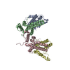
|
|---|---|
| 1 | x 60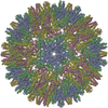
|
| 2 |
|
| 3 | x 5
|
| 4 | x 6
|
| 5 | 
|
| Symmetry | Point symmetry: (Schoenflies symbol: I (icosahedral)) |
-Components
| #1: Protein | Mass: 21398.465 Da / Num. of mol.: 4 / Fragment: UNP residues 30-214 Source method: isolated from a genetically manipulated source Source: (gene. exp.) Hepatitis B virus / Gene: C, PreC / Production host:  |
|---|
-Experimental details
-Experiment
| Experiment | Method: ELECTRON MICROSCOPY |
|---|---|
| EM experiment | Aggregation state: PARTICLE / 3D reconstruction method: single particle reconstruction |
- Sample preparation
| Component | Name: Hepatitis B virus core / Type: VIRUS |
|---|---|
| Specimen | Embedding applied: NO / Shadowing applied: NO / Staining applied: NO / Vitrification applied: YES |
| Vitrification | Instrument: FEI VITROBOT MARK II / Cryogen name: NITROGEN |
- Electron microscopy imaging
| Experimental equipment |  Model: Titan Krios / Image courtesy: FEI Company |
|---|---|
| Microscopy | Model: FEI TITAN KRIOS / Date: Jan 1, 2009 |
| Electron gun | Electron source:  FIELD EMISSION GUN / Accelerating voltage: 300 kV / Illumination mode: FLOOD BEAM FIELD EMISSION GUN / Accelerating voltage: 300 kV / Illumination mode: FLOOD BEAM |
| Electron lens | Mode: BRIGHT FIELD / Nominal magnification: 75000 X |
| Image recording | Electron dose: 25 e/Å2 |
| Radiation | Protocol: SINGLE WAVELENGTH / Monochromatic (M) / Laue (L): M / Scattering type: x-ray |
| Radiation wavelength | Relative weight: 1 |
- Processing
| Symmetry | Point symmetry: I (icosahedral) | ||||||||||||
|---|---|---|---|---|---|---|---|---|---|---|---|---|---|
| 3D reconstruction | Resolution: 3.5 Å / Num. of particles: 8093 / Symmetry type: POINT | ||||||||||||
| Refinement step | Cycle: LAST
|
