 Movie
Movie Controller


+ Open data

- Basic information
| Entry | Database: PDB / ID: 3j15 | ||||||
|---|---|---|---|---|---|---|---|
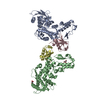
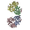
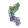
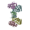
| Title | Model of ribosome-bound archaeal Pelota and ABCE1 | ||||||
 Components Components |
| ||||||
 Keywords Keywords | TRANSLATION/TRANSPORT PROTEIN / ribosome recycling /  ribosome / archaea / TRANSLATION-TRANSPORT PROTEIN complex ribosome / archaea / TRANSLATION-TRANSPORT PROTEIN complex | ||||||
| Function / homology |  Function and homology information Function and homology informationRNA surveillance / nuclear-transcribed mRNA catabolic process, no-go decay / nuclear-transcribed mRNA catabolic process, non-stop decay / nonfunctional rRNA decay / ribosome disassembly / ribosome binding / endonuclease activity / Hydrolases; Acting on ester bonds / metal ion binding / cytoplasmSimilarity search - Function | ||||||
| Biological species |   Thermococcus kodakarensis (archaea)Pyrococcus furiosus (archaea) Thermococcus kodakarensis (archaea)Pyrococcus furiosus (archaea) | ||||||
| Method | ELECTRON MICROSCOPY / single particle reconstruction / cryo EM / Resolution: 6.6 Å | ||||||
 Authors Authors | Becker, T. / Franckenberg, S. / Wickles, S. / Shoemaker, C.J. / Anger, A.M. / Armache, J.-P. / Sieber, H. / Ungewickell, C. / Berninghausen, O. / Daberkow, I. ...Becker, T. / Franckenberg, S. / Wickles, S. / Shoemaker, C.J. / Anger, A.M. / Armache, J.-P. / Sieber, H. / Ungewickell, C. / Berninghausen, O. / Daberkow, I. / Karcher, A. / Thomm, M. / Hopfner, K.-P. / Green, R. / Beckmann, R. | ||||||
 Citation Citation | Journal: Nature / Year: 2012 Title: Structural basis of highly conserved ribosome recycling in eukaryotes and archaea. Authors: Thomas Becker / Sibylle Franckenberg / Stephan Wickles / Christopher J Shoemaker / Andreas M Anger / Jean-Paul Armache / Heidemarie Sieber / Charlotte Ungewickell / Otto Berninghausen / Ingo ...Authors: Thomas Becker / Sibylle Franckenberg / Stephan Wickles / Christopher J Shoemaker / Andreas M Anger / Jean-Paul Armache / Heidemarie Sieber / Charlotte Ungewickell / Otto Berninghausen / Ingo Daberkow / Annette Karcher / Michael Thomm / Karl-Peter Hopfner / Rachel Green / Roland Beckmann /  Abstract: Ribosome-driven protein biosynthesis is comprised of four phases: initiation, elongation, termination and recycling. In bacteria, ribosome recycling requires ribosome recycling factor and elongation ...Ribosome-driven protein biosynthesis is comprised of four phases: initiation, elongation, termination and recycling. In bacteria, ribosome recycling requires ribosome recycling factor and elongation factor G, and several structures of bacterial recycling complexes have been determined. In the eukaryotic and archaeal kingdoms, however, recycling involves the ABC-type ATPase ABCE1 and little is known about its structural basis. Here we present cryo-electron microscopy reconstructions of eukaryotic and archaeal ribosome recycling complexes containing ABCE1 and the termination factor paralogue Pelota. These structures reveal the overall binding mode of ABCE1 to be similar to canonical translation factors. Moreover, the iron-sulphur cluster domain of ABCE1 interacts with and stabilizes Pelota in a conformation that reaches towards the peptidyl transferase centre, thus explaining how ABCE1 may stimulate peptide-release activity of canonical termination factors. Using the mechanochemical properties of ABCE1, a conserved mechanism in archaea and eukaryotes is suggested that couples translation termination to recycling, and eventually to re-initiation. | ||||||
| History |
|
- Structure visualization
| Movie |
Movie viewer |
|---|---|
| Structure viewer | Molecule: MolmilJmol/JSmol |
- Downloads & links
-Download
| PDBx/mmCIF format | 3j15.cif.gz | 176.9 KB | Display | PDBx/mmCIF format |
|---|---|---|---|---|
| PDB format | pdb3j15.ent.gz | 140.3 KB | Display | PDB format |
| PDBx/mmJSON format | 3j15.json.gz | Tree view | PDBx/mmJSON format | |
| Others |  Other downloads Other downloads |
-Validation report
| Arichive directory | https://data.pdbj.org/pub/pdb/validation_reports/j1/3j15ftp://data.pdbj.org/pub/pdb/validation_reports/j1/3j15 | HTTPS FTP |
|---|
-Related structure data
| Related structure data |  2009MC 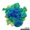 2008C  2010C 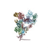 3j16C C: citing same article ( M: map data used to model this data |
|---|---|
| Similar structure data |


-Links
 PDBj
PDBj
- Assembly
| Deposited unit | 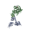
|
|---|---|
| 1 |
|
-Components
| #1: Protein | Mass: 40673.898 Da / Num. of mol.: 1 Source method: isolated from a genetically manipulated source Source: (gene. exp.) Thermococcus kodakarensis (archaea) / Gene: pelA, TK0964 / Plasmid: pET28 / Production host:  Escherichia coli BL21(DE3) (bacteria) / Strain (production host): Rosetta(DE3) Escherichia coli BL21(DE3) (bacteria) / Strain (production host): Rosetta(DE3)References: UniProt: Q5JIB9, Hydrolases; Acting on ester bonds | ||||
|---|---|---|---|---|---|
| #2: Protein | Mass: 67286.273 Da / Num. of mol.: 1 Source method: isolated from a genetically manipulated source Source: (gene. exp.) Pyrococcus furiosus (archaea) / Plasmid: pET28 / Production host: Escherichia coli BL21(DE3) (bacteria) / Strain (production host): Rosetta(DE3) | ||||
| #3: Chemical | Adenosine diphosphate  Mass: 427.201 Da / Num. of mol.: 2 / Source method: obtained synthetically / Formula: C10H15N5O10P2 / Comment: ADP, energy-carrying molecule*YM Mass: 427.201 Da / Num. of mol.: 2 / Source method: obtained synthetically / Formula: C10H15N5O10P2 / Comment: ADP, energy-carrying molecule*YM#4: Chemical | Iron–sulfur cluster  Mass: 351.640 Da / Num. of mol.: 2 / Source method: obtained synthetically / Formula: Fe4S4 Mass: 351.640 Da / Num. of mol.: 2 / Source method: obtained synthetically / Formula: Fe4S4Sequence details | MODELED SEQUENCES WERE DERIVED FROM PYROCOCCUS KODAKARAENSIS (PELOTA) AND PYROCOCCUS ABYSSI (ABCE1). ...MODELED SEQUENCES WERE DERIVED FROM PYROCOCCUS | |
-Experimental details
-Experiment
| Experiment | Method: ELECTRON MICROSCOPY |
|---|---|
| EM experiment | Aggregation state: PARTICLE / 3D reconstruction method: single particle reconstruction |
- Sample preparation
| Component | Name: 70S ABCE1-Pelota complex / Type: RIBOSOME |
|---|---|
| Buffer solution | pH: 8.2 |
| Specimen | Embedding applied: NO / Shadowing applied: NO / Staining applied: NO / Vitrification applied: YES |
| Vitrification | Instrument: FEI VITROBOT MARK I / Cryogen name: ETHANE / Humidity: 95 % Details: Blot for 10 seconds before plunging into ethane (Vitrobot) Method: Blot for 10 seconds before plunging, use 2 layer of filter paper |
- Electron microscopy imaging
| Experimental equipment |  Model: Titan Krios / Image courtesy: FEI Company |
|---|---|
| Microscopy | Model: FEI TITAN KRIOS |
| Electron gun | Electron source: FIELD EMISSION GUN / Accelerating voltage: 200 kV / Illumination mode: FLOOD BEAM |
| Electron lens | Mode: BRIGHT FIELDBright-field microscopy / Nominal magnification: 75000 X / Nominal defocus max: 3600 nm / Nominal defocus min: 1200 nm |
| Image recording | Electron dose: 25 e/Å2 / Film or detector model: TVIPS TEMCAM-F416 (4k x 4k) |
- Processing
| EM software | Name: SPIDER / Category: 3D reconstruction | ||||||||||||
|---|---|---|---|---|---|---|---|---|---|---|---|---|---|
| Symmetry | Point symmetry: C1 (asymmetric) | ||||||||||||
| 3D reconstruction | Method: Single particleSingle particle analysis / Resolution: 6.6 Å / Num. of particles: 51000 / Nominal pixel size: 1.2375 Å / Actual pixel size: 1.2375 Å / Symmetry type: POINT | ||||||||||||
| Refinement step | Cycle: LAST
|
