 Movie
Movie Controller

[English] 日本語
 Yorodumi
- PDB-3j0r: Model of a type III secretion system needle based on a 7 Angstrom... -
+ Open data

- Basic information
| Entry | Database: PDB / ID: 3j0r | ||||||
|---|---|---|---|---|---|---|---|
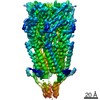
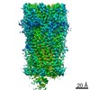
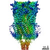
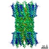
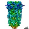
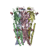
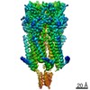
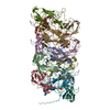
| Title | Model of a type III secretion system needle based on a 7 Angstrom resolution cryoEM map | ||||||
 Components Components | Protein mxiH | ||||||
 Keywords Keywords | PROTEIN TRANSPORT / helical assembly | ||||||
| Function / homology |  Function and homology information Function and homology informationtype III protein secretion system complex / protein secretion by the type III secretion system / cell surface / extracellular region / identical protein binding Similarity search - Function | ||||||
| Biological species |  Shigella flexneri (bacteria) Shigella flexneri (bacteria) | ||||||
| Method | ELECTRON MICROSCOPY / helical reconstruction / cryo EM / Resolution: 7.7 Å | ||||||
 Authors Authors | Fujii, T. / Cheung, M. / Blanco, A. / Kato, T. / Blocker, A.J. / Namba, K. | ||||||
 Citation Citation | Journal: Proc Natl Acad Sci U S A / Year: 2012 Title: Structure of a type III secretion needle at 7-Å resolution provides insights into its assembly and signaling mechanisms. Authors: Takashi Fujii / Martin Cheung / Amandine Blanco / Takayuki Kato / Ariel J Blocker / Keiichi Namba /  Abstract: Type III secretion systems of Gram-negative bacteria form injection devices that deliver effector proteins into eukaryotic cells during infection. They span both bacterial membranes and the ...Type III secretion systems of Gram-negative bacteria form injection devices that deliver effector proteins into eukaryotic cells during infection. They span both bacterial membranes and the extracellular space to connect with the host cell plasma membrane. Their extracellular portion is a needle-like, hollow tube that serves as a secretion conduit for effector proteins. The needle of Shigella flexneri is approximately 50-nm long and 7-nm thick and is made by the helical assembly of one protein, MxiH. We provide a 7-Å resolution 3D image reconstruction of the Shigella needle by electron cryomicroscopy, which resolves α-helices and a β-hairpin that has never been observed in the crystal and solution structures of needle proteins, including MxiH. An atomic model of the needle based on the 3D-density map, in comparison with that of the bacterial-flagellar filament, provides insights into how such a thin tubular structure is stably assembled by intricate intermolecular interactions. The map also illuminates how the needle-length control protein functions as a ruler within the central channel during export of MxiH for assembly at the distal end of the needle, and how the secretion-activation signal may be transduced through a conformational change of the needle upon host-cell contact. | ||||||
| History |
|
- Structure visualization
| Movie |
Movie viewer |
|---|---|
| Structure viewer | Molecule: MolmilJmol/JSmol |
- Downloads & links
-Download
| PDBx/mmCIF format | 3j0r.cif.gz | 24.9 KB | Display | PDBx/mmCIF format |
|---|---|---|---|---|
| PDB format | pdb3j0r.ent.gz | 15.4 KB | Display | PDB format |
| PDBx/mmJSON format | 3j0r.json.gz | Tree view | PDBx/mmJSON format | |
| Others |  Other downloads Other downloads |
-Validation report
| Summary document | 3j0r_validation.pdf.gz | 862.8 KB | Display | wwPDB validaton report |
|---|---|---|---|---|
| Full document | 3j0r_full_validation.pdf.gz | 875.8 KB | Display | |
| Data in XML | 3j0r_validation.xml.gz | 12.3 KB | Display | |
| Data in CIF | 3j0r_validation.cif.gz | 15.7 KB | Display | |
| Arichive directory | https://data.pdbj.org/pub/pdb/validation_reports/j0/3j0rftp://data.pdbj.org/pub/pdb/validation_reports/j0/3j0r | HTTPS FTP |
-Related structure data
| Related structure data | 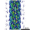 5352MC M: map data used to model this data C: citing same article ( |
|---|---|
| Similar structure data |
-Links
 PDBj
PDBj- Assembly
| Deposited unit | 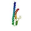
|
|---|---|
| 1 | x 22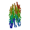
|
| 2 |
|
| 3 | 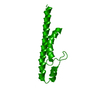
|
| Symmetry | Helical symmetry: (Circular symmetry: 1 / Dyad axis: no / N subunits divisor: 1 / Num. of operations: 22 / Rise per n subunits: 4.3 Å / Rotation per n subunits: 64.06 °) |
-Components
| #1: Protein | Mass: 9141.068 Da / Num. of mol.: 1 / Source method: isolated from a natural source / Source: (natural) Shigella flexneri (bacteria) / References: UniProt: P0A223 |
|---|
-Experimental details
-Experiment
| Experiment | Method: ELECTRON MICROSCOPY |
|---|---|
| EM experiment | Aggregation state: FILAMENT / 3D reconstruction method: helical reconstruction |
- Sample preparation
| Component | Name: type III secretion needle / Type: COMPLEX / Details: a helical assembly of MxiH |
|---|---|
| Buffer solution | Name: 20 mM Tris, pH 7.4, 150 mM NaCl, 2mM MgSO4 / pH: 7.4 / Details: 20 mM Tris, pH 7.4, 150 mM NaCl, 2mM MgSO4 |
| Specimen | Embedding applied: NO / Shadowing applied: NO / Staining applied: NO / Vitrification applied: YES |
| Specimen support | Details: Quantifoil holey carbon molybdenum grid (R0.6/1.0, Quantifoil) |
| Vitrification | Instrument: FEI VITROBOT MARK I / Cryogen name: ETHANE Details: Blot for 3.5 seconds before plunging into liquid ethane (Vitrobot). |
- Electron microscopy imaging
| Microscopy | Model: JEOL 3200FSC / Date: Jul 2, 2008 |
|---|---|
| Electron gun | Electron source:  FIELD EMISSION GUN / Accelerating voltage: 200 kV / Illumination mode: FLOOD BEAM FIELD EMISSION GUN / Accelerating voltage: 200 kV / Illumination mode: FLOOD BEAM |
| Electron lens | Mode: BRIGHT FIELD / Nominal magnification: 50000 X / Calibrated magnification: 89285 X / Nominal defocus max: 2000 nm / Nominal defocus min: 1000 nm / Cs: 1.6 mm |
| Specimen holder | Temperature: 50 K |
| Image recording | Electron dose: 20 e/Å2 / Film or detector model: TVIPS TEMCAM-F415 (4k x 4k) |
- Processing
| CTF correction | Details: CTFFIND3 Each particle | ||||||||||||
|---|---|---|---|---|---|---|---|---|---|---|---|---|---|
| Helical symmerty | Angular rotation/subunit: 64.06 ° / Axial rise/subunit: 4.3 Å / Axial symmetry: C1 | ||||||||||||
| 3D reconstruction | Method: Projection Matching / Resolution: 7.7 Å / Symmetry type: HELICAL | ||||||||||||
| Atomic model building | Space: REAL | ||||||||||||
| Refinement step | Cycle: LAST
|
