 Movie
Movie Controller

[English] 日本語
 Yorodumi
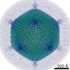
- PDB-3iyl: Atomic CryoEM Structure of a Nonenveloped Virus Suggests How Memb... -
+ Open data

- Basic information
| Entry | Database: PDB / ID: 3iyl | ||||||
|---|---|---|---|---|---|---|---|
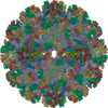
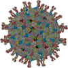
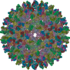
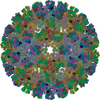
| Title | Atomic CryoEM Structure of a Nonenveloped Virus Suggests How Membrane Penetration Protein is Primed for Cell Entry | ||||||
 Components Components |
| ||||||
 Keywords Keywords |  VIRUS / Non-enveloped virus / Membrane penetration protein / Autocleavage / Myristol Group / Icosahedral virus VIRUS / Non-enveloped virus / Membrane penetration protein / Autocleavage / Myristol Group / Icosahedral virus | ||||||
| Function / homology |  Function and homology information Function and homology informationhost cell surface binding / viral inner capsid / viral outer capsid / permeabilization of host organelle membrane involved in viral entry into host cell / symbiont entry into host cell via permeabilization of inner membrane / 7-methylguanosine mRNA capping / viral capsid / mRNA guanylyltransferase activity / mRNA 5'-cap (guanine-N7-)-methyltransferase activity / RNA helicase activity ...host cell surface binding / viral inner capsid / viral outer capsid / permeabilization of host organelle membrane involved in viral entry into host cell / symbiont entry into host cell via permeabilization of inner membrane / 7-methylguanosine mRNA capping / viral capsid / mRNA guanylyltransferase activity / mRNA 5'-cap (guanine-N7-)-methyltransferase activity / RNA helicase activity / RNA helicase / hydrolase activity / GTP binding / ATP binding / metal ion bindingSimilarity search - Function | ||||||
| Biological species |  Grass carp reovirus Grass carp reovirus | ||||||
| Method | ELECTRON MICROSCOPY / single particle reconstruction / cryo EM / Resolution: 3.3 Å | ||||||
 Authors Authors | Zhang, X. / Jin, L. / Fang, Q. / Hui, W. / Zhou, Z.H. | ||||||
 Citation Citation | Journal: Cell / Year: 2010 Title: 3.3 A cryo-EM structure of a nonenveloped virus reveals a priming mechanism for cell entry. Authors: Xing Zhang / Lei Jin / Qin Fang / Wong H Hui / Z Hong Zhou /  Abstract: To achieve cell entry, many nonenveloped viruses must transform from a dormant to a primed state. In contrast to the membrane fusion mechanism of enveloped viruses (e.g., influenza virus), this ...To achieve cell entry, many nonenveloped viruses must transform from a dormant to a primed state. In contrast to the membrane fusion mechanism of enveloped viruses (e.g., influenza virus), this membrane penetration mechanism is poorly understood. Here, using single-particle cryo-electron microscopy, we report a 3.3 A structure of the primed, infectious subvirion particle of aquareovirus. The density map reveals side-chain densities of all types of amino acids (except glycine), enabling construction of a full-atom model of the viral particle. Our structure and biochemical results show that priming involves autocleavage of the membrane penetration protein and suggest that Lys84 and Glu76 may facilitate this autocleavage in a nucleophilic attack. We observe a myristoyl group, covalently linked to the N terminus of the penetration protein and embedded in a hydrophobic pocket. These results suggest a well-orchestrated process of nonenveloped virus entry involving autocleavage of the penetration protein prior to exposure of its membrane-insertion finger. | ||||||
| History |
|
- Structure visualization
| Movie |
Movie viewer |
|---|---|
| Structure viewer | Molecule: MolmilJmol/JSmol |
- Downloads & links
-Download
| PDBx/mmCIF format | 3iyl.cif.gz | 1.8 MB | Display | PDBx/mmCIF format |
|---|---|---|---|---|
| PDB format | pdb3iyl.ent.gz | 1.5 MB | Display | PDB format |
| PDBx/mmJSON format | 3iyl.json.gz | Tree view | PDBx/mmJSON format | |
| Others |  Other downloads Other downloads |
-Validation report
| Arichive directory | https://data.pdbj.org/pub/pdb/validation_reports/iy/3iylftp://data.pdbj.org/pub/pdb/validation_reports/iy/3iyl | HTTPS FTP |
|---|
-Related structure data
| Related structure data | 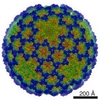 5160MC M: map data used to model this data C: citing same article ( |
|---|---|
| Similar structure data |

-Links
 PDBj
PDBj


- Assembly
| Deposited unit | 
|
|---|---|
| 1 | x 60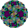
|
| 2 |
|
| 3 | x 5
|
| 4 | x 6
|
| 5 | 
|
| Symmetry | Point symmetry: (Schoenflies symbol: I (icosahedral)) |
-Components
| #1: Protein | Mass: 68646.750 Da / Num. of mol.: 20 / Source method: isolated from a natural source / Source: (natural) Grass carp reovirus / References: UniProt: Q8JU67#2: Protein | Mass: 44606.535 Da / Num. of mol.: 2 / Source method: isolated from a natural source / Source: (natural) Grass carp reovirus / References: UniProt: Q8JU64#3: Protein | | Mass: 141512.156 Da / Num. of mol.: 1 / Source method: isolated from a natural source / Source: (natural) Grass carp reovirus / References: UniProt: Q9E3W0#4: Protein | Mass: 132203.312 Da / Num. of mol.: 2 / Source method: isolated from a natural source / Source: (natural) Grass carp reovirus / References: UniProt: Q9E3V8#5: Chemical | ChemComp-MYR / Myristic acid  Mass: 228.371 Da / Num. of mol.: 10 / Source method: obtained synthetically / Formula: C14H28O2 Mass: 228.371 Da / Num. of mol.: 10 / Source method: obtained synthetically / Formula: C14H28O2 |
|---|
-Experimental details
-Experiment
| Experiment | Method: ELECTRON MICROSCOPY |
|---|---|
| EM experiment | Aggregation state: PARTICLE / 3D reconstruction method: single particle reconstruction |
- Sample preparation
| Component | Name: Aquareovirus / Type: VIRUS / Details: The sample was monodisperse |
|---|---|
| Molecular weight | Value: 72 MDa / Experimental value: NO |
| Details of virus | Empty: NO / Enveloped: NO / Host category: VERTEBRATES / Isolate: STRAIN / Type: VIRION |
| Buffer solution | pH: 7.5 / Details: 10mM PBS Buffer |
| Specimen | Embedding applied: NO / Shadowing applied: NO / Staining applied: NO / Vitrification applied: YES / Details: 10mM PBS Buffer |
| Vitrification | Instrument: FEI VITROBOT MARK I / Cryogen name: METHANE / Temp: 90 K / Humidity: 100 % / Method: Blot for 7-9 seconds before plunging |
- Electron microscopy imaging
| Experimental equipment |  Model: Titan Krios / Image courtesy: FEI Company |
|---|---|
| Microscopy | Model: FEI TITAN KRIOS / Date: Mar 1, 2009 |
| Electron gun | Electron source: FIELD EMISSION GUN / Accelerating voltage: 300 kV / Illumination mode: FLOOD BEAM |
| Electron lens | Mode: BRIGHT FIELDBright-field microscopy / Nominal magnification: 59000 X / Calibrated magnification: 57700 X / Nominal defocus max: 2700 nm / Nominal defocus min: 400 nm / Cs: 2.7 mm Astigmatism : objective lens astigmatism was corrected at 250,000 times magnificationCamera length: 0 mm |
| Specimen holder | Specimen holder model: OTHER / Specimen holder type: Eucentric / Temperature: 90 K / Temperature (min): 90 K / Tilt angle max: 0 ° / Tilt angle min: 0 ° |
| Image recording | Electron dose: 25 e/Å2 / Film or detector model: KODAK SO-163 FILM |
| Radiation | Protocol: SINGLE WAVELENGTH / Monochromatic (M) / Laue (L): M |
| Radiation wavelength | Relative weight: 1 |
- Processing
| EM software |
| ||||||||||||
|---|---|---|---|---|---|---|---|---|---|---|---|---|---|
| CTF correction | Details: Each particle | ||||||||||||
| Symmetry | Point symmetry: I (icosahedral) | ||||||||||||
| 3D reconstruction | Method: Fourier Space Reconstruction / Resolution: 3.3 Å / Resolution method: FSC 0.143 CUT-OFF / Num. of particles: 18464 / Symmetry type: POINT | ||||||||||||
| Refinement step | Cycle: LAST
|
