[English] 日本語
Yorodumi
- PDB-3foi: Fitting of gp18M crystal structure into 3D cryo-EM reconstruction... -

+
Open data


ID or keywords:

Loading...

-
Basic information

Entry
Database: PDB / ID: 3foi
TitleFitting of gp18M crystal structure into 3D cryo-EM reconstruction of bacteriophage T4 contracted tail
ComponentsTail sheath protein Gp18
KeywordsVIRAL PROTEIN / alpha-beta / viral structural protein / bacteriophage T4 / tail sheath
Function / homologyvirus tail, sheath / Phage tail sheath protein, beta-sandwich domain / Phage tail sheath protein beta-sandwich domain / symbiont genome ejection through host cell envelope, contractile tail mechanism / Tail sheath protein, subtilisin-like domain / Phage tail sheath protein subtilisin-like domain / Tail sheath protein, C-terminal domain / Phage tail sheath C-terminal domain / Tail sheath protein
Function and homology information
Biological speciesEnterobacteria phage T4 (virus)
MethodELECTRON MICROSCOPY / single particle reconstruction / cryo EM / Resolution: 16 Å
AuthorsAksyuk, A.A. / Leiman, P.G. / Kurochkina, L.P. / Shneider, M.M. / Kostyuchenko, V.A. / Mesyanzhinov, V.V. / Rossmann, M.G.
Citation
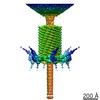
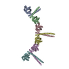
Journal: EMBO J / Year: 2009
Title: The tail sheath structure of bacteriophage T4: a molecular machine for infecting bacteria.
Authors: Anastasia A Aksyuk / Petr G Leiman / Lidia P Kurochkina / Mikhail M Shneider / Victor A Kostyuchenko / Vadim V Mesyanzhinov / Michael G Rossmann /
Abstract: The contractile tail of bacteriophage T4 is a molecular machine that facilitates very high viral infection efficiency. Its major component is a tail sheath, which contracts during infection to less ...The contractile tail of bacteriophage T4 is a molecular machine that facilitates very high viral infection efficiency. Its major component is a tail sheath, which contracts during infection to less than half of its initial length. The sheath consists of 138 copies of the tail sheath protein, gene product (gp) 18, which surrounds the central non-contractile tail tube. The contraction of the sheath drives the tail tube through the outer membrane, creating a channel for the viral genome delivery. A crystal structure of about three quarters of gp18 has been determined and was fitted into cryo-electron microscopy reconstructions of the tail sheath before and after contraction. It was shown that during contraction, gp18 subunits slide over each other with no apparent change in their structure.
History
DepositionDec 30, 2008Deposition site: RCSB / Processing site: RCSB
Revision 1.0Mar 10, 2009Provider: repository / Type: Initial release
Revision 1.1Jul 13, 2011Group: Version format compliance
Revision 1.2May 30, 2012Group: Refinement description
Revision 1.3Jul 18, 2018Group: Data collection / Category: em_image_scans / em_software / Item: _em_software.image_processing_id
Revision 1.4Nov 6, 2019Group: Data collection / Other / Category: atom_sites / cell
Item: _atom_sites.fract_transf_matrix[1][1] / _atom_sites.fract_transf_matrix[2][2] ..._atom_sites.fract_transf_matrix[1][1] / _atom_sites.fract_transf_matrix[2][2] / _atom_sites.fract_transf_matrix[3][3] / _cell.Z_PDB / _cell.length_a / _cell.length_b / _cell.length_c
Revision 1.5Oct 20, 2021Group: Database references / Category: database_2 / struct_ref_seq_dif
Item: _database_2.pdbx_DOI / _database_2.pdbx_database_accession / _struct_ref_seq_dif.details
Revision 1.6Feb 21, 2024Group: Data collection / Refinement description
Category: chem_comp_atom / chem_comp_bond ...chem_comp_atom / chem_comp_bond / em_3d_fitting_list / pdbx_initial_refinement_model
Item: _em_3d_fitting_list.accession_code / _em_3d_fitting_list.initial_refinement_model_id ..._em_3d_fitting_list.accession_code / _em_3d_fitting_list.initial_refinement_model_id / _em_3d_fitting_list.source_name / _em_3d_fitting_list.type

-
Structure visualization

Movie
  • Deposited structure unit
  • Imaged by Jmol
  • Download
  • Simplified surface model + fitted atomic model
  • EMDB-1086
  • Imaged by Jmol
  • Download
Movie viewer
Structure viewerMolecule:
MolmilJmol/JSmol

Downloads & links

-
Assembly

Deposited unit
A: Tail sheath protein Gp18
B: Tail sheath protein Gp18
C: Tail sheath protein Gp18
D: Tail sheath protein Gp18
E: Tail sheath protein Gp18
F: Tail sheath protein Gp18


Theoretical massNumber of molelcules
Total (without water)327,8766
Polymers327,8766
Non-polymers00
Water0
1


  • Idetical with deposited unit
  • defined by author
TypeNameSymmetry operationNumber
identity operation1_555x,y,z1

-
Components

#1: Protein
Tail sheath protein Gp18


Mass: 54645.965 Da / Num. of mol.: 6 / Fragment: deletion mutant gp18M: UNP Residues 1-510 / Mutation: R510P
Source method: isolated from a genetically manipulated source
Details: six gp18M fitted into one disk of the tail sheath in the contracted conformation
Source: (gene. exp.) Enterobacteria phage T4 (virus) / Gene: 18, GB AAA32541 / Plasmid: pET22 / Production host: Escherichia coli (E. coli) / Strain (production host): BL21(DE3) / References: UniProt: P13332
Sequence detailsAUTHORS STATE THAT THE SEQUENCE COMPLETELY AGREES WITH THE GENBANK ENTRY AAA32541, PUBMED REPORT ...AUTHORS STATE THAT THE SEQUENCE COMPLETELY AGREES WITH THE GENBANK ENTRY AAA32541, PUBMED REPORT 2964531. THE SEQUENCE DIFFERENCES MAY REFLECT THE SEQUENCE VARIATION BETWEEN THE VIRUS STRAINS.

-
Experimental details

-
Experiment

ExperimentMethod: ELECTRON MICROSCOPY
EM experimentAggregation state: PARTICLE / 3D reconstruction method: single particle reconstruction

-
Sample preparation

Component
IDNameTypeParent-IDDetails
1bacteriophage T4 extended tailVIRUS0
2one disk of the tail sheath in the contracted conformation1Six gp18 subunits form one disk of the tail sheath. 23 disks form a bacteriophage T4 tail. During infection the tail sheath subunits change their relative positions resulting in the contraction of the tail sheath.
Buffer solutionpH: 7.5 / Details: H2O
SpecimenEmbedding applied: NO / Shadowing applied: NO / Staining applied: NO / Vitrification applied: YES
VitrificationCryogen name: ETHANE

-
Electron microscopy imaging

MicroscopyModel: FEI/PHILIPS CM300FEG/T / Date: Jan 6, 2002
Electron gunElectron source: FIELD EMISSION GUN / Accelerating voltage: 300 kV / Illumination mode: SPOT SCAN
Electron lensMode: BRIGHT FIELDBright-field microscopy / Nominal magnification: 45000 X / Nominal defocus max: 3400 nm / Nominal defocus min: 500 nm
Image recordingElectron dose: 20 e/Å2 / Film or detector model: KODAK SO-163 FILM

-
Processing

EM software
IDNameCategory
1SITUS COLORESmodel fitting
2SPIDER3D reconstruction
SymmetryPoint symmetry: C6 (6 fold cyclic)
3D reconstructionResolution: 16 Å / Num. of particles: 1965 / Symmetry type: POINT
Atomic model buildingPDB-ID: 3FOA
Accession code: 3FOA / Source name: PDB / Type: experimental model
Refinement stepCycle: LAST
ProteinNucleic acidLigandSolventTotal
Num. atoms21678 0 0 0 21678

+
About Yorodumi

-
News

-
Feb 9, 2022. New format data for meta-information of EMDB entries

New format data for meta-information of EMDB entries

  • Version 3 of the EMDB header file is now the official format.
  • The previous official version 1.9 will be removed from the archive.

Related info.:EMDB header

External links:wwPDB to switch to version 3 of the EMDB data model

-
Aug 12, 2020. Covid-19 info

Covid-19 info

URL: https://pdbj.org/emnavi/covid19.php

New page: Covid-19 featured information page in EM Navigator.

Related info.:Covid-19 info / Mar 5, 2020. Novel coronavirus structure data

+
Mar 5, 2020. Novel coronavirus structure data

Novel coronavirus structure data

Related info.:Yorodumi Speices / Aug 12, 2020. Covid-19 info

External links:COVID-19 featured content - PDBj / Molecule of the Month (242):Coronavirus Proteases

+
Jan 31, 2019. EMDB accession codes are about to change! (news from PDBe EMDB page)

EMDB accession codes are about to change! (news from PDBe EMDB page)

  • The allocation of 4 digits for EMDB accession codes will soon come to an end. Whilst these codes will remain in use, new EMDB accession codes will include an additional digit and will expand incrementally as the available range of codes is exhausted. The current 4-digit format prefixed with “EMD-” (i.e. EMD-XXXX) will advance to a 5-digit format (i.e. EMD-XXXXX), and so on. It is currently estimated that the 4-digit codes will be depleted around Spring 2019, at which point the 5-digit format will come into force.
  • The EM Navigator/Yorodumi systems omit the EMD- prefix.

Related info.:Q: What is EMD? / ID/Accession-code notation in Yorodumi/EM Navigator

External links:EMDB Accession Codes are Changing Soon! / Contact to PDBj

+
Jul 12, 2017. Major update of PDB

Major update of PDB

  • wwPDB released updated PDB data conforming to the new PDBx/mmCIF dictionary.
  • This is a major update changing the version number from 4 to 5, and with Remediation, in which all the entries are updated.
  • In this update, many items about electron microscopy experimental information are reorganized (e.g. em_software).
  • Now, EM Navigator and Yorodumi are based on the updated data.

External links:wwPDB Remediation / Enriched Model Files Conforming to OneDep Data Standards Now Available in the PDB FTP Archive

-
Yorodumi

Thousand views of thousand structures

  • Yorodumi is a browser for structure data from EMDB, PDB, SASBDB, etc.
  • This page is also the successor to EM Navigator detail page, and also detail information page/front-end page for Omokage search.
  • The word "yorodu" (or yorozu) is an old Japanese word meaning "ten thousand". "mi" (miru) is to see.

Related info.:EMDB / PDB / SASBDB / Comparison of 3 databanks / Yorodumi Search / Aug 31, 2016. New EM Navigator & Yorodumi / Yorodumi Papers / Jmol/JSmol / Function and homology information / Changes in new EM Navigator and Yorodumi

Read more