 Movie
Movie Controller


+ Open data

- Basic information
| Entry | Database: PDB / ID: 2z9q | ||||||
|---|---|---|---|---|---|---|---|
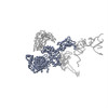
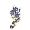
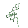
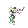
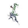
| Title | Transfer RNA in the hybrid P/E state | ||||||
 Components Components | tRNA | ||||||
 Keywords Keywords | RNA / distorted anticodon-stem-loop / twisted CCA arm | ||||||
| Function / homology | RNA / RNA (> 10) Function and homology information Function and homology information | ||||||
| Biological species |   Thermus aquaticus (bacteria) Thermus aquaticus (bacteria) | ||||||
| Method | ELECTRON MICROSCOPY / single particle reconstruction / cryo EM / Resolution: 11.7 Å | ||||||
 Authors Authors | Frank, J. / Li, W. | ||||||
 Citation Citation | Journal: Proc Natl Acad Sci U S A / Year: 2007 Title: Transfer RNA in the hybrid P/E state: correlating molecular dynamics simulations with cryo-EM data. Authors: Wen Li / Joachim Frank /  Abstract: Transfer RNA (tRNA) transiently occupies the hybrid P/E state (P/E-tRNA) when mRNA-tRNA are translocated in the ribosome. In this study, we characterize the structure of P/E-tRNA and its interactions ...Transfer RNA (tRNA) transiently occupies the hybrid P/E state (P/E-tRNA) when mRNA-tRNA are translocated in the ribosome. In this study, we characterize the structure of P/E-tRNA and its interactions with the ribosome by correlating the results from molecular dynamics simulations on free tRNA with the cryo-EM map of P/E-tRNA. In our approach, we show that the cryo-EM map may be interpreted as a conformational average. Along the molecular dynamics trajectories (44 ns, 18 ns, and 18 ns), some of the snapshots prove to be quite close to the observed density. In a representative structure, the CCA (3') arm is uniquely twisted, and the anticodon stem loop is kinked at the junctions to both the anticodon loop and the D stem. In addition, the map shows that the P/E-tRNA is no longer bound to helix H69 of 23S rRNA and is flexible, and the conformations of helices H68 and h44 of 16S rRNA differ from those in the x-ray structure. Thus, our study presents structural and dynamic information on the P/E-tRNA and characterizes its interactions with the translocating ribosome. #1: Journal: Cell / Year: 2003Title: Locking and unlocking of ribosomal motions. Authors: Mikel Valle / Andrey Zavialov / Jayati Sengupta / Urmila Rawat / Måns Ehrenberg / Joachim Frank / Abstract: During the ribosomal translocation, the binding of elongation factor G (EF-G) to the pretranslocational ribosome leads to a ratchet-like rotation of the 30S subunit relative to the 50S subunit in the ...During the ribosomal translocation, the binding of elongation factor G (EF-G) to the pretranslocational ribosome leads to a ratchet-like rotation of the 30S subunit relative to the 50S subunit in the direction of the mRNA movement. By means of cryo-electron microscopy we observe that this rotation is accompanied by a 20 A movement of the L1 stalk of the 50S subunit, implying that this region is involved in the translocation of deacylated tRNAs from the P to the E site. These ribosomal motions can occur only when the P-site tRNA is deacylated. Prior to peptidyl-transfer to the A-site tRNA or peptide removal, the presence of the charged P-site tRNA locks the ribosome and prohibits both of these motions. | ||||||
| History |
|
- Structure visualization
| Movie |
Movie viewer |
|---|---|
| Structure viewer | Molecule: MolmilJmol/JSmol |
- Downloads & links
-Download
| PDBx/mmCIF format | 2z9q.cif.gz | 15.8 KB | Display | PDBx/mmCIF format |
|---|---|---|---|---|
| PDB format | pdb2z9q.ent.gz | 4.6 KB | Display | PDB format |
| PDBx/mmJSON format | 2z9q.json.gz | Tree view | PDBx/mmJSON format | |
| Others |  Other downloads Other downloads |
-Validation report
| Summary document | 2z9q_validation.pdf.gz | 746.2 KB | Display | wwPDB validaton report |
|---|---|---|---|---|
| Full document | 2z9q_full_validation.pdf.gz | 745.9 KB | Display | |
| Data in XML | 2z9q_validation.xml.gz | 8.5 KB | Display | |
| Data in CIF | 2z9q_validation.cif.gz | 10.6 KB | Display | |
| Arichive directory | https://data.pdbj.org/pub/pdb/validation_reports/z9/2z9qftp://data.pdbj.org/pub/pdb/validation_reports/z9/2z9q | HTTPS FTP |
-Related structure data
| Related structure data | 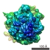 1363M M: map data used to model this data |
|---|---|
| Similar structure data |


-Links
 PDBj
PDBj


- Assembly
| Deposited unit | 
|
|---|---|
| 1 |
|
-Components
| #1: RNA chain | Mass: 24544.916 Da / Num. of mol.: 1 / Source method: isolated from a natural source / Source: (natural) Thermus aquaticus (bacteria) |
|---|
-Experimental details
-Experiment
| Experiment | Method: ELECTRON MICROSCOPY |
|---|---|
| EM experiment | Aggregation state: PARTICLE / 3D reconstruction method: single particle reconstruction |
- Sample preparation
| Component | Name: EF-G bound Release Complex in the presence of Puromycin and GDPNP Type: RIBOSOME |
|---|---|
| Buffer solution | Name: polymix / pH: 7.5 / Details: polymix |
| Specimen | Conc.: 32 mg/ml / Embedding applied: NO / Shadowing applied: NO / Staining applied: NO / Vitrification applied: YES |
| Specimen support | Details: carbon on quantifoil grids |
| Vitrification | Cryogen name: ETHANE / Details: PLUNGED INTO ETHANE |
- Electron microscopy imaging
| Experimental equipment |  Model: Tecnai F20 / Image courtesy: FEI Company |
|---|---|
| Microscopy | Model: FEI TECNAI F20 / Date: Jun 1, 2001 / Details: SAMPLES WERE MAINTAINED AT LIQUID NITROGEN |
| Electron gun | Electron source:  FIELD EMISSION GUN / Accelerating voltage: 200 kV / Illumination mode: FLOOD BEAM FIELD EMISSION GUN / Accelerating voltage: 200 kV / Illumination mode: FLOOD BEAM |
| Electron lens | Mode: BRIGHT FIELD / Nominal magnification: 50000 X / Calibrated magnification: 49696 X / Nominal defocus max: 3.925 nm / Nominal defocus min: 1.75 nm / Cs: 2 mm |
| Specimen holder | Temperature: 80 K / Tilt angle max: 0 ° / Tilt angle min: 0 ° |
| Image recording | Electron dose: 15 e/Å2 / Film or detector model: KODAK SO-163 FILM |
| Image scans | Num. digital images: 38858 |
| Radiation | Protocol: SINGLE WAVELENGTH / Monochromatic (M) / Laue (L): M / Scattering type: x-ray |
| Radiation wavelength | Relative weight: 1 |
- Processing
| EM software | Name: SPIDER / Category: 3D reconstruction Details: Fast Motif Search Procedure in SPIDER. Reference: Rath and Frank 2004 JSB 145 page84 | ||||||||||||
|---|---|---|---|---|---|---|---|---|---|---|---|---|---|
| CTF correction | Details: CTF correction of each defocus group reconstruction | ||||||||||||
| Symmetry | Point symmetry: C1 (asymmetric) | ||||||||||||
| 3D reconstruction | Method: single particle reconstruction / Resolution: 11.7 Å / Nominal pixel size: 2.8 Å / Actual pixel size: 2.82 Å / Magnification calibration: TMV / Details: THE STRUCTURE CONTAINS P ATOMS ONLY / Symmetry type: POINT | ||||||||||||
| Atomic model building | Protocol: OTHER / Space: REAL / Target criteria: cross correlation coefficient / Details: REFINEMENT PROTOCOL--auto | ||||||||||||
| Atomic model building | PDB-ID: 1TTT Accession code: 1TTT / Source name: PDB / Type: experimental model | ||||||||||||
| Refinement step | Cycle: LAST /
|
